. 2015 Oct 27;10(10):e0141043.
doi: 10.1371/journal.pone.0141043. eCollection 2015.

In Situ Localization and Rhythmic Expression of Ghrelin and ghs-r1 Ghrelin Receptor in the Brain and Gastrointestinal Tract of Goldfish (Carassius auratus)

Affiliations

In Situ Localization and Rhythmic Expression of Ghrelin and ghs-r1 Ghrelin Receptor in the Brain and Gastrointestinal Tract of Goldfish (Carassius auratus)

Aída Sánchez-Bretaño et al. PLoS One. .

Abstract

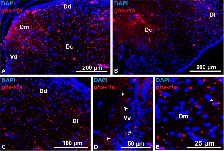
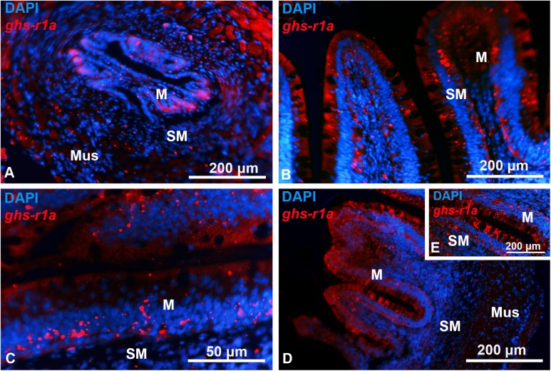
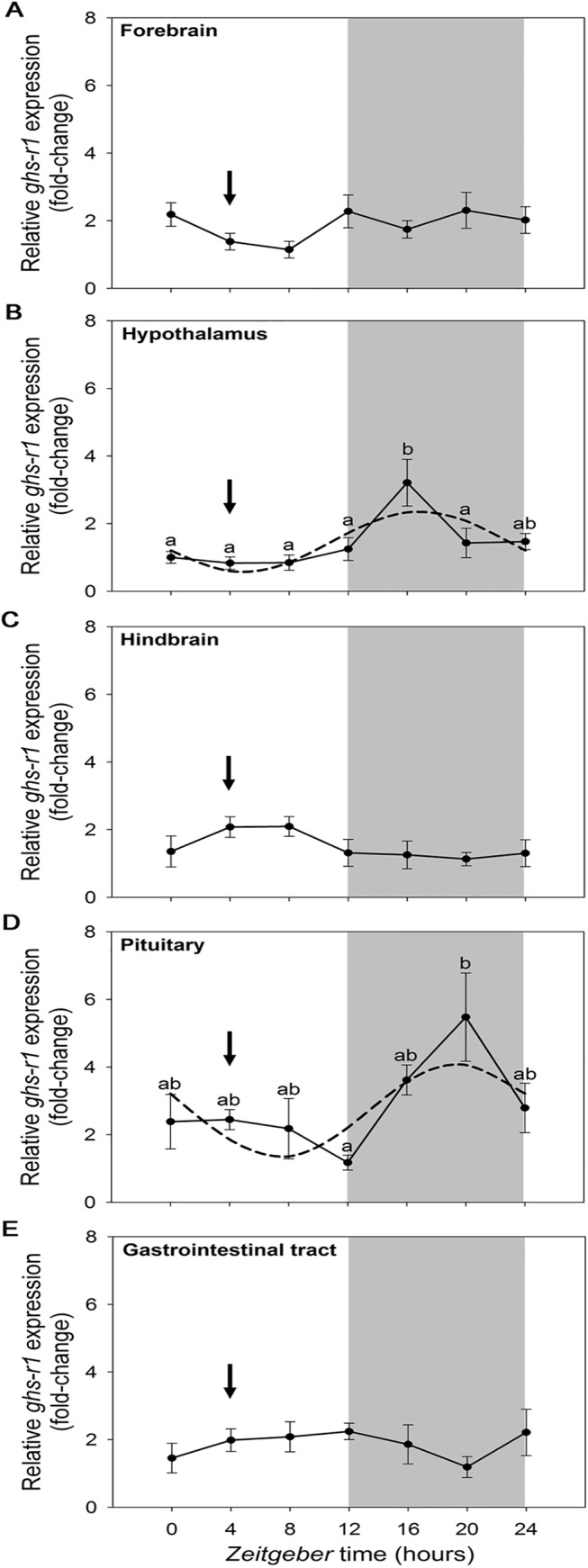
Ghrelin is a gut-brain peptide hormone, which binds to the growth hormone secretagogue receptor (GHS-R) to regulate a wide variety of biological processes in fish. Despite these prominent physiological roles, no studies have reported the anatomical distribution of preproghrelin transcripts using in situ hybridization in a non-mammalian vertebrate, and its mapping within the different encephalic areas remains unknown. Similarly, no information is available on the possible 24-h variations in the expression of preproghrelin and its receptor in any vertebrate species. The first aim of this study was to investigate the anatomical distribution of ghrelin and GHS-R1a ghrelin receptor subtype in brain and gastrointestinal tract of goldfish (Carassius auratus) using immunohistochemistry and in situ hybridization. Our second aim was to characterize possible daily variations of preproghrelin and ghs-r1 mRNA expression in central and peripheral tissues using real-time reverse transcription-quantitative PCR. Results show ghrelin expression and immunoreactivity in the gastrointestinal tract, with the most abundant signal observed in the mucosal epithelium. These are in agreement with previous findings on mucosal cells as the primary synthesizing site of ghrelin in goldfish. Ghrelin receptor was observed mainly in the hypothalamus with low expression in telencephalon, pineal and cerebellum, and in the same gastrointestinal areas as ghrelin. Daily rhythms in mRNA expression were found for preproghrelin and ghs-r1 in hypothalamus and pituitary with the acrophase occurring at nighttime. Preproghrelin, but not ghs-r1a, displayed a similar daily expression rhythm in the gastrointestinal tract with an amplitude 3-fold higher than the rest of tissues. Together, these results described for the first time in fish the mapping of preproghrelin and ghrelin receptor ghs-r1a in brain and gastrointestinal tract of goldfish, and provide the first evidence for a daily regulation of both genes expression in such locations, suggesting a possible connection between the ghrelinergic and circadian systems in teleosts.


Conflict of interest statement

Competing Interests: The authors have declared that no competing interests exist.

Figures

Fig 1
Fig 1. Transversal representative sections of goldfish gastrointestinal tract showing preproghrelin positive cells identified by in situ hybridization (left panel) and ghrelin immunoreactive cells detected by immunohistochemistry (right panel).
(A, B) Esophagus. (C, D) Intestinal bulb. (E, F) j-loop. (G, H) Anterior intestine. M, mucose; Mus, muscular layer; SM, submucose. Scale bars are indicated in each image.
Fig 2
Fig 2. Schematic representation of ghs-r1a expressing cells in antero-posterior (A to F) transversal sections of goldfish brain [Carassius auratus Forebrain Atlas [43]].
The brain areas shown in the transversal sections are indicated with a schematic representation in the upper part of each image. Intensity of signal is represented by density of red dots. Dc, central portion of the dorsal telencephalon; Dd, dorsal portion of the dorsal telencephalon; Dl, lateral portion of the dorsal telencephalon; Dld, dorsal part of the lateral portion of the dorsal telencephalon; Dlv ventral part of the lateral portion of the dorsal telencephalon; Dm, medial portion of the dorsal telencephalon; NAPv, anterior periventricular nucleus; NH habenular nucleus; NPO, preoptic nucleus; NPP, periventricular preoptic nucleus; NRL, lateral recess nucleus; NRP, posterior recess nucleus; OT, optic tract; P, pineal; TL, torus longitudinalis; Vc, valvula of the cerebellum; Vd, dorsal portion of the ventral telencephalon; Vl, lateral portion of the ventral telencephalon; Vp, postcommissural portion of the ventral telencephalon; Vs, supracommisural portion of the ventral telencephalon; Vv, ventral portion of the ventral telencephalon.
Fig 3
Fig 3. Transversal representative sections of goldfish telencephalon showing ghs-r1a positive cells detected by in situ hybridization.
(A, B) Overview of telencephalon. (C) Lateral portion of the dorsal telencephalon. (D) Ventral portion of the ventral telencephalon (arrowheads indicate riboprobe signaling). (E) Example of telencephalic nucleus surrounded by ghs-r1a mRNA riboprobe (arrowhead). Dc, central portion of the dorsal telencephalon; Dd, dorsal portion of the dorsal telencephalon; Dl, lateral portion of the dorsal telencephalon; Dm, medial portion of the dorsal telencephalon; Vd, dorsal portion of the ventral telencephalon; Vv, ventral portion of the ventral telencephalon. Scale bars are indicated in each image.
Fig 4
Fig 4. Transversal representative sections of goldfish hypothalamus showing ghs-r1a positive cells detected by in situ hybridization.
(A) Periventricular preoptic nucleus. (B) Preoptic recess. (C) Preoptic nucleus. (D) Posterior recess nucleus. E, F, G. Lateral recess nucleus. NAPv, anterior periventricular nucleus; NPO, preoptic nucleus; NPP, periventricular preoptic nucleus; NRL, lateral recess nucleus; NRP, posterior recess nucleus; OT, optic tract. Scale bars are indicated in each image.
Fig 5
Fig 5. Transversal representative sections of goldfish pineal (A), habenula (B), torus longitudinalis (C) and valvula of the cerebellum (D) showing ghs-r1a positive cells detected by in situ hybridization.
NH habenular nucleus; P, pineal; TL, torus longitudinalis; Vc, valvula cerebelli. Scale bars are indicated in each image.
Fig 6
Fig 6. Transversal representative sections of goldfish gastrointestinal tract showing ghs-r1a positive cells detected by in situ hybridization.
(A) Esophagus. (B) Intestinal bulb. (C) j-loop. (D, E) Anterior intestine. M, mucose; Mus, muscular layer; SM, submucose. Scale bars are indicated in each image.
Fig 7
Fig 7. Relative expression of preproghrelin in goldfish forebrain (A), hypothalamus (B), hindbrain (C), pituitary (D) and gastrointestinal tract (E) during a 24-h light/dark cycle.
Relative mRNA amounts were quantified by RT-qPCR. Data are expressed as mean ± SEM (n = 6/time point). The grey area indicates the dark phase of the daily photocycle, and the arrow indicates the scheduled feeding time (ZT-4). Dashed lines represent the periodic sinusoidal functions determined by the cosinor analysis when a significant rhythm was detected. Different letters indicate significant differences by ANOVA and post-hoc SNK test (p<0.05).
Fig 8
Fig 8. Relative expression of ghs-r1 in goldfish forebrain (A), hypothalamus (B), hindbrain (C), pituitary (D) and gastrointestinal tract (E) during a 24-h light/dark cycle.
Relative mRNA amounts were quantified by RT-qPCR. Data are expressed as mean ± SEM (n = 6/time point). The grey area indicates the dark phase of the daily photocycle, and the arrow indicates the scheduled feeding time (ZT-4). Dashed lines represent the periodic sinusoidal functions determined by the cosinor analysis when a significant rhythm was detected. Different letters indicate significant differences by ANOVA and post-hoc SNK test (p<0.05).

References

    1. Kojima M, Hosoda H, Date Y, Nakazato M, Matsuo H, Kangawa K. Ghrelin is a growth-hormone-releasing acylated peptide from stomach. Nature. 1999; 402: 656–660. - PubMed
    1. Kojima M, Kangawa K. Ghrelin: structure and function. Physiol Rev. 2005; 85: 495–522. - PubMed
    1. Gnanapavan S, Kola B, Bustin SA, Morris DG, McGee P, Fairclough P, et al. The tissue distribution of the mRNA of ghrelin and subtypes of its receptor, GHS-R, in humans. J Clin Endocrinol Metab. 2002; 87: 2988–2991. - PubMed
    1. Ghelardoni S, Carnicelli V, Frascarelli S, Ronca-Testoni S, Zucchi R. Ghrelin tissue distribution: comparison between gene and protein expression. J Endocrinol Invest. 2006; 29: 115–121. - PubMed
    1. Unniappan S, Lin X, Cervini L, Rivier J, Kaiya H, Kangawa K, et al. Goldfish ghrelin: molecular characterization of the complementary deoxyribonucleic acid, partial gene structure and evidence for its stimulatory role in food intake. Endocrinology. 2002; 143: 4143–4146. - PubMed

Publication types

LinkOut - more resources