Review
. 2016 Feb 27;428(5 Pt B):963-89.
doi: 10.1016/j.jmb.2015.10.014. Epub 2015 Oct 23.

Origins of Programmable Nucleases for Genome Engineering

Affiliations
Review

Origins of Programmable Nucleases for Genome Engineering

Srinivasan Chandrasegaran et al. J Mol Biol. .

Abstract

Genome engineering with programmable nucleases depends on cellular responses to a targeted double-strand break (DSB). The first truly targetable reagents were the zinc finger nucleases (ZFNs) showing that arbitrary DNA sequences could be addressed for cleavage by protein engineering, ushering in the breakthrough in genome manipulation. ZFNs resulted from basic research on zinc finger proteins and the FokI restriction enzyme (which revealed a bipartite structure with a separable DNA-binding domain and a non-specific cleavage domain). Studies on the mechanism of cleavage by 3-finger ZFNs established that the preferred substrates were paired binding sites, which doubled the size of the target sequence recognition from 9 to 18bp, long enough to specify a unique genomic locus in plant and mammalian cells. Soon afterwards, a ZFN-induced DSB was shown to stimulate homologous recombination in cells. Transcription activator-like effector nucleases (TALENs) that are based on bacterial TALEs fused to the FokI cleavage domain expanded this capability. The fact that ZFNs and TALENs have been used for genome modification of more than 40 different organisms and cell types attests to the success of protein engineering. The most recent technology platform for delivering a targeted DSB to cellular genomes is that of the RNA-guided nucleases, which are based on the naturally occurring Type II prokaryotic CRISPR-Cas9 system. Unlike ZFNs and TALENs that use protein motifs for DNA sequence recognition, CRISPR-Cas9 depends on RNA-DNA recognition. The advantages of the CRISPR-Cas9 system-the ease of RNA design for new targets and the dependence on a single, constant Cas9 protein-have led to its wide adoption by research laboratories around the world. These technology platforms have equipped scientists with an unprecedented ability to modify cells and organisms almost at will, with wide-ranging implications across biology and medicine. However, these nucleases have also been shown to cut at off-target sites with mutagenic consequences. Therefore, issues such as efficacy, specificity and delivery are likely to drive selection of reagents for particular purposes. Human therapeutic applications of these technologies will ultimately depend on risk versus benefit analysis and informed consent.

Keywords: CRISPR-Cas9; gene therapy; genome editing; transcription activator-like effector nucleases (TALENs); zinc finger nucleases (ZFNs).


Figures

Fig. 1
Fig. 1
Genome engineering using programmable nucleases. ZFNs, TALENs and CRISPR-Cas9 are used to induce a targeted DSB at the desired chromosomal locus. Either non-homologous end joining (NHEJ) or homology-directed repair (HDR), one of the two cellular repair pathways, is then used to repair the DSB. Error-prone NHEJ could be used to knockout genes, while HDR could be used for either gene correction or to introduce precise alterations into the genome directed by an investigator-provided homologous DNA template. Figure adapted from Ramalingam et al 2013.
Fig. 2
Fig. 2
Publication milestones for genome engineering using programmable nucleases.
Fig. 3
Fig. 3
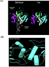
Crystal structure of FokI and FokI bound to DNA. (A), Structure of FokI-DNA complex and FokI enzyme alone. In both structures, the FokI cleavage domain piggybacks on the recognition domain. (B). Native FokI crystallizes as a dimer. The dimer interface is at the FokI nuclease domains, which is formed by two salt bridges between arginine (R) and aspartic acid (D) residues of the FokI monomers. Figure adapted from Wah et al 1998.
Fig. 4
Fig. 4
DNA recognition by ZFPs. (A), Structure of ZFs; (B), DNA recognition by ZFs; and (C) Structure of 3-finger Zif268 bound to its cognate site. Figure adapted from Pabo et al 2001 & Miller and Pabo, 2001.
Fig. 5
Fig. 5
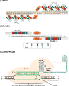
A schematic diagram depicting programmable nucleases recognizing their target sites. (A), ZFNs; (B), TALENs and (C), RNA-guided CRISPR-Cas9. Figure adapted from Ramalingam et al 2013.
Fig. 6
Fig. 6
TALEN-mediated generation of Cystic fibrosis (CF) and Gaucher’s disease (GD) human induced pluripotent stem cells (hiPSCs) from patient-derived fibroblasts. (A), Schematic diagram depicts the two-step protocol that was used to generate GD hiPSCs. In a first step, the donor containing five stem cell factor genes (OSKLN: Oct4, Sox2, Klf4, Lin8 and Nanog) and eGFP, was inserted at the safe harbor CCR5 locus (using CCR5-specific TALENS) to reprogram the patient-derived fibroblasts. In a second step, the loxP site-flanked donor was excised from the CCR5-modified CF and GD hiPSCs by treatment with Cre reombinase. CCR5-specific ZFNs have also been used similarly to generate of hiPSCs from human fibroblasts and cord blood cells. (B) Bright field images of the morphology of CF and GD hiPSCs generated using CCR5-specific TALENs. Characterization of GD hiPSCs by Oct4/Sox2/nanog/Tra-1–60 immunostaining and DAPI staining are also shown. (C) TALEN-mediated correction of SCD hiPSCs. Monoallele correction of homozygous HBB mutation of patient-specific SCD hiPSCs [TNC1 line] was achieved using HBB-specific TALENs and wild-type HBB donor construct. Monoallele gene correction was confirmed by sequencing the HBB locus. Parts of figure adapted from Ramalingam et al 2014.
Fig. 6
Fig. 6
TALEN-mediated generation of Cystic fibrosis (CF) and Gaucher’s disease (GD) human induced pluripotent stem cells (hiPSCs) from patient-derived fibroblasts. (A), Schematic diagram depicts the two-step protocol that was used to generate GD hiPSCs. In a first step, the donor containing five stem cell factor genes (OSKLN: Oct4, Sox2, Klf4, Lin8 and Nanog) and eGFP, was inserted at the safe harbor CCR5 locus (using CCR5-specific TALENS) to reprogram the patient-derived fibroblasts. In a second step, the loxP site-flanked donor was excised from the CCR5-modified CF and GD hiPSCs by treatment with Cre reombinase. CCR5-specific ZFNs have also been used similarly to generate of hiPSCs from human fibroblasts and cord blood cells. (B) Bright field images of the morphology of CF and GD hiPSCs generated using CCR5-specific TALENs. Characterization of GD hiPSCs by Oct4/Sox2/nanog/Tra-1–60 immunostaining and DAPI staining are also shown. (C) TALEN-mediated correction of SCD hiPSCs. Monoallele correction of homozygous HBB mutation of patient-specific SCD hiPSCs [TNC1 line] was achieved using HBB-specific TALENs and wild-type HBB donor construct. Monoallele gene correction was confirmed by sequencing the HBB locus. Parts of figure adapted from Ramalingam et al 2014.

Comment in

References

    1. Mansour SL, Thomas KR, Capecchi MR. Disruption of the proto-oncogene int-2 in mouse embryo-derived stem cells: a general strategy for targeting mutations to non-selectable genes. Nature. 1988;336(6197):348–352. - PubMed
    1. Capecchi MR. Altering the genome by homologous recombination. Science. 1989;244:1288–1292. - PubMed
    1. Rouet P, Smih, Jasin M. Introduction of double-strand breaks into the genome of mouse cells by expression of a rare-cutting endonuclease. Mol Cell Biol. 1994;14(12):8096–8106. - PMC - PubMed
    1. Pingoud A, Wilson GG, Wende W. Type II restriction endonuclease - - a historical perspective and more. Nucl Acids Res. 2014;42:7489–727. - PMC - PubMed
    1. Kita K, Kotani H, Sugisaki H, Takanami M. The FokI restriction-modification systems. I Organization and nucleotide sequences of the restriction and modification genes. J Biol Chem. 1989;264:5751–5756. - PubMed

Publication types