. 2015 Oct 27;6(6):e01428-15.
doi: 10.1128/mBio.01428-15.

Toxoplasma gondii Infections Alter GABAergic Synapses and Signaling in the Central Nervous System

Affiliations

Toxoplasma gondii Infections Alter GABAergic Synapses and Signaling in the Central Nervous System

Justin M Brooks et al. mBio. .

Abstract

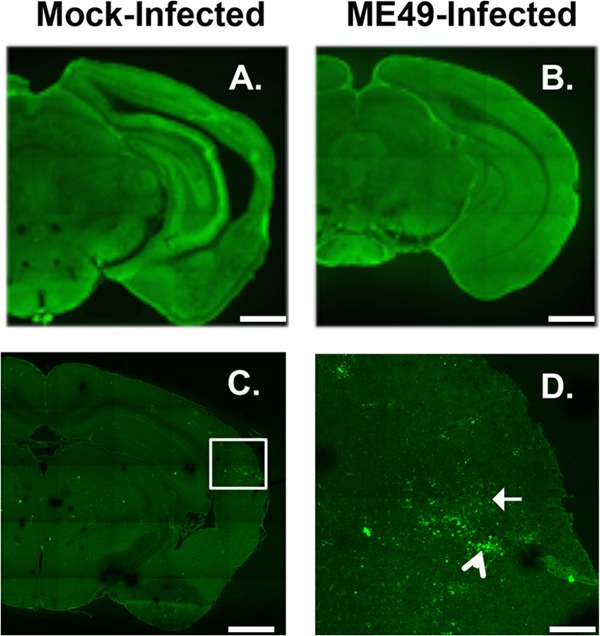
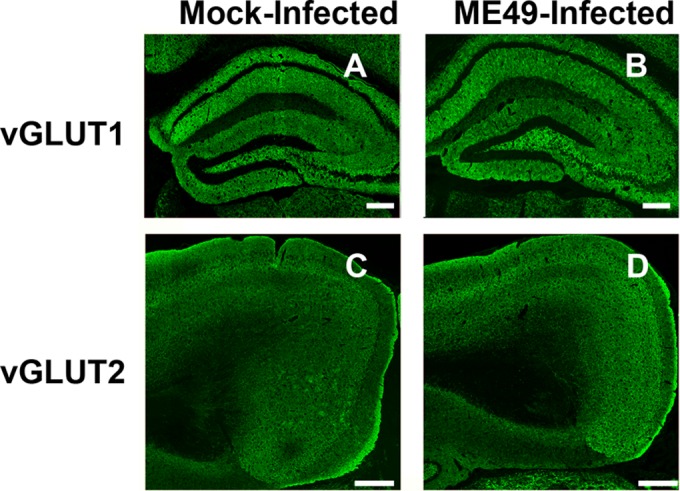
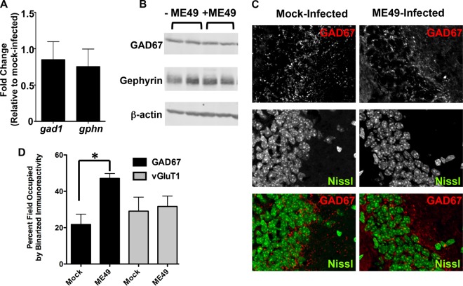
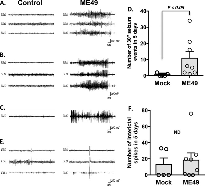
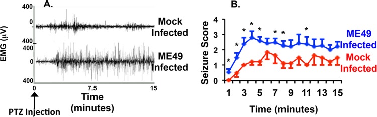
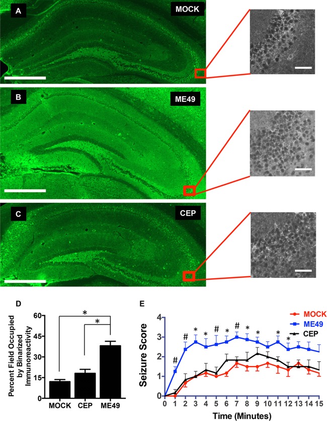
During infections with the protozoan parasite Toxoplasma gondii, gamma-aminobutyric acid (GABA) is utilized as a carbon source for parasite metabolism and also to facilitate parasite dissemination by stimulating dendritic-cell motility. The best-recognized function for GABA, however, is its role in the nervous system as an inhibitory neurotransmitter that regulates the flow and timing of excitatory neurotransmission. When this pathway is altered, seizures develop. Human toxoplasmosis patients suffer from seizures, suggesting that Toxoplasma interferes with GABA signaling in the brain. Here, we show that while excitatory glutamatergic presynaptic proteins appeared normal, infection with type II ME49 Toxoplasma tissue cysts led to global changes in the distribution of glutamic acid decarboxylase 67 (GAD67), a key enzyme that catalyzes GABA synthesis in the brain. Alterations in GAD67 staining were not due to decreased expression but rather to a change from GAD67 clustering at presynaptic termini to a more diffuse localization throughout the neuropil. Consistent with a loss of GAD67 from the synaptic terminals, Toxoplasma-infected mice develop spontaneous seizures and are more susceptible to drugs that induce seizures by antagonizing GABA receptors. Interestingly, GABAergic protein mislocalization and the response to seizure-inducing drugs were observed in mice infected with type II ME49 but not type III CEP strain parasites, indicating a role for a polymorphic parasite factor(s) in regulating GABAergic synapses. Taken together, these data support a model in which seizures and other neurological complications seen in Toxoplasma-infected individuals are due, at least in part, to changes in GABAergic signaling.

Importance: Infections of the central nervous system can cause seizures. While inflammation in the brain has been proposed to initiate the onset of the seizures, relatively little is known about how inflammation impacts the structure and function of the neurons. Here we used a parasite called Toxoplasma gondii that infects the brain and showed that seizures arise due to a defect in signaling of GABA, which is the neurotransmitter primarily responsible for preventing the onset of seizures.


Figures

FIG 1
FIG 1
Decreased GABAergic synaptic protein staining in type II ME49 Toxoplasma-infected brains. (A and B) GAD67 immunoreactivity in mock-infected (A) or type II ME49 Toxoplasma-infected (B) brains. (C and D) Detection of type II ME49 Toxoplasma tachyzoites (white arrow in panel D) and bradyzoites (arrowhead in panel D). The image in panel D is a high-magnification image of the boxed in area in panel C. Scale bars, 1 mm (A, B, and C) and 0.25 mm (D). Lines appear in the images due to lack of overlap during image acquisition of each brain.
FIG 2
FIG 2
Unaltered glutamatergic synaptic protein staining in type II ME49 Toxoplasma-infected brains. (A and B) VGluT1 immunoreactivity in mock-infected or type II ME49 Toxoplasma-infected hippocampi. Scale bars, 0.2 mm. (C and D) VGluT2 immunoreactivity in mock-infected or type II ME49 Toxoplasma-infected cortexes. Scale bars, 0.4 mm.
FIG 3
FIG 3
Type II ME49 Toxoplasma infections induce mislocalization of GAD67. (A) RT-PCR quantification of relative gad1 and gphn transcript levels in mock-infected and type II ME49 parasite-infected brains. (B) Western blot detection of GAD67, gephyrin, and β-actin in cortical tissues harvested from mock-infected and type II ME49 parasite-infected brains. (C) High-magnification images of GAD67 staining in hippocampal CA3 regions in mock-infected and type II ME49 parasite-infected mice. Note the change from punctate staining in the mock-infected brains to a more diffuse staining in the parasite-infected brains. (D) Quantification of percent GAD67 and VGlut1 staining following binarizing of 5 images/brain (n = 3 mice/group). *, P < 0.02 (unpaired, two-tailed t test).
FIG 4
FIG 4
Type II ME49 Toxoplasma infection leads to spontaneous seizures in mice. (A and B) Examples of EEG and EMG recordings from control (left column) and type II ME49-infected (right column) mice. Data represent 2 EEG traces and 1 EMG trace that were obtained simultaneously from each animal and are shown in a time-locked state. The recordings represented in panel A show 60 s of neural and muscular activity in control and type II ME49-infected mice. The recordings represented in panel B show 120 s of neural and muscular activity in control and type II ME49-infected mice. (C) EMG recordings from control (left column) and type II ME49-infected (right column) mice. Note that motor seizures persisted for as long as 15 min in this type II ME49-infected mouse. (D) Quantification of the number of seizure events that exceeded 30 s in the 5-day recording period. Bar graph data represent means ± SEM. Circles represent individual data points. P < 0.05 (Tukey-Kramer HSD test). (E) Individual and sparse spikes of neural and muscular activity were observed at similar frequencies in mock-infected and type II ME49-infected mice. (F) Quantification of the number of spiking events in the 5-day recording period. Bar graph data represent means ± SEM. Circles represent individual data points. The numbers of spikes seen with mock-infected mice were not statistically significantly different from those seen with type II ME49-infected mice (P > 0.1 [Tukey-Kramer HSD test]).
FIG 5
FIG 5
Increased susceptibility of type II ME49 Toxoplasma-infected mice to drug-induced seizures. (A) Five days after mice were analyzed for spontaneous seizures, they were injected with PTZ (40 mg/kg), and EEG/EMG data were recorded for 15 min. The data shown represent typical EMG traces from a mock-infected mouse (top) and a type II ME49 parasite-infected mouse (bottom). Notice the increased strength and intensity of muscular activity in the type II ME49 Toxoplasma-infected mouse. (B) Averages (± SEM) of seizure score data recorded every minute for 15 min after injection of PTZ. n = 5 mice for wild-type mice and 9 for type II ME49 mice. *, P < 0.05 (unpaired, two-tailed t test).
FIG 6
FIG 6
GAD67 mislocalization and drug-induced seizure are polymorphic responses in Toxoplasma-infected brains. (A to C) Representative images of coronal sections from mock-infected (A), type II ME49-infected (B), and type III CEP-infected (C) mice 30 days postinfection. Scale bars, 500 µm. Expanded images show high-magnification views of hippocampus CA3 regions. Scale bars, 50 µm. (D) Quantification of percent GAD67 staining following binarizing of images. *, P < 0.001 (one-way ANOVA with Tukey’s post hoc test; n ≥ 3 mice for each group). (E) Averages (± SEM) of seizure scores of mock-infected, type II ME49-infected, and type III CEP-infected mice (30 days postinfection) recorded every minute for 15 min after injection of PTZ. Each group represents score data for a minimum of 6 mice. *, statistically significant difference between type II ME49-infected and mock-infected mice only (P < 0.05 [one-way ANOVA with Tukey’s post hoc test]). #, statistically significant difference between type II ME49 and both mock-infected and type III CEP-infected mice (P < 0.05 [one-way ANOVA with Tukey’s post hoc test]).

References

    1. Xu D, Miller SD, Koh S. 2013. Immune mechanisms in epileptogenesis. Front Cell Neurosci 7:195. doi: 10.3389/fncel.2013.00195. - DOI - PMC - PubMed
    1. Annegers JF, Hauser WA, Coan SP, Rocca WA. 1998. A population-based study of seizures after traumatic brain injuries. N Engl J Med 338:20–24. doi: 10.1056/NEJM199801013380104. - DOI - PubMed
    1. Avila EK, Graber J. 2010. Seizures and epilepsy in cancer patients. Curr Neurol Neurosci Rep 10:60–67. doi: 10.1007/s11910-009-0080-z. - DOI - PubMed
    1. Marchi N, Angelov L, Masaryk T, Fazio V, Granata T, Hernandez N, Hallene K, Diglaw T, Franic L, Najm I, Janigro D. 2007. Seizure-promoting effect of blood-brain barrier disruption. Epilepsia 48:732–742. doi: 10.1111/j.1528-1167.2007.00988.x. - DOI - PMC - PubMed
    1. Vezzani A, Maroso M, Balosso S, Sanchez M, Bartfai T. 2011. IL-1 receptor/Toll-like receptor signaling in infection, inflammation, stress and neurodegeneration couples hyperexcitability and seizures. Brain Behav Immun 25:1281–1289. doi: 10.1016/j.bbi.2011.03.018. - DOI - PubMed

Substances

LinkOut - more resources