

North Carolina Macular Dystrophy Is Caused by Dysregulation of the Retinal Transcription Factor PRDM13
- PMID: 26507665
- PMCID: PMC4695238
- DOI: 10.1016/j.ophtha.2015.10.006
North Carolina Macular Dystrophy Is Caused by Dysregulation of the Retinal Transcription Factor PRDM13
Abstract
Purpose: To identify specific mutations causing North Carolina macular dystrophy (NCMD).
Design: Whole-genome sequencing coupled with reverse transcription polymerase chain reaction (RT-PCR) analysis of gene expression in human retinal cells.
Participants: A total of 141 members of 12 families with NCMD and 261 unrelated control individuals.
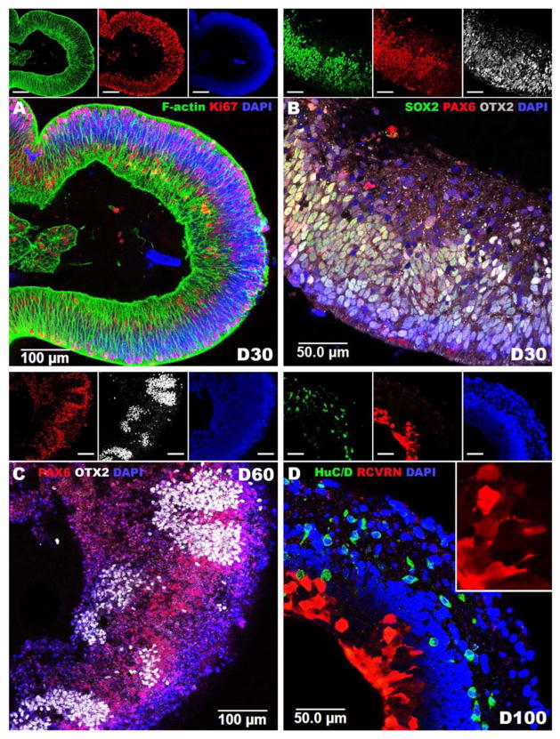
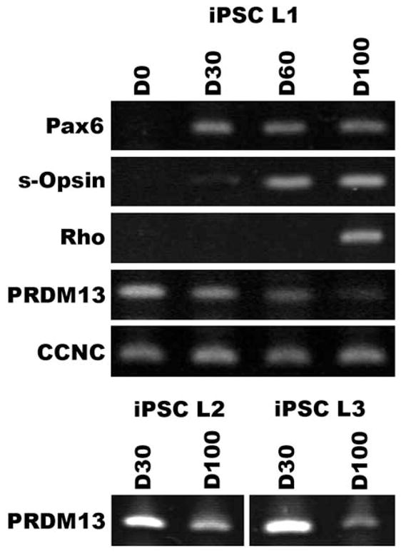
Methods: Genome sequencing was performed on 8 affected individuals from 3 families affected with chromosome 6-linked NCMD (MCDR1) and 2 individuals affected with chromosome 5-linked NCMD (MCDR3). Variants observed in the MCDR1 locus with frequencies <1% in published databases were confirmed using Sanger sequencing. Confirmed variants absent from all published databases were sought in 8 additional MCDR1 families and 261 controls. The RT-PCR analysis of selected genes was performed in stem cell-derived human retinal cells.
Main outcome measures: Co-segregation of rare genetic variants with disease phenotype.
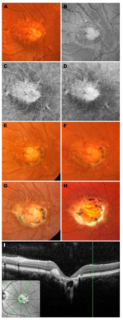
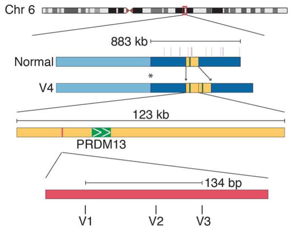
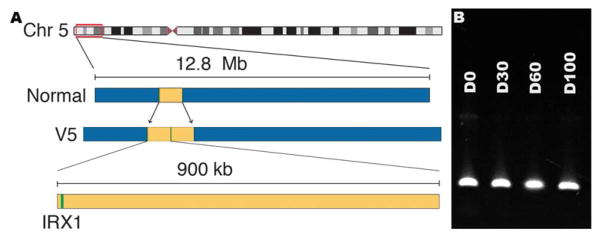
Results: Five sequenced individuals with MCDR1-linked NCMD shared a haplotype of 14 rare variants spanning 1 Mb of the disease-causing allele. One of these variants (V1) was absent from all published databases and all 261 controls, but was found in 5 additional NCMD kindreds. This variant lies in a DNase 1 hypersensitivity site (DHS) upstream of both the PRDM13 and CCNC genes. Sanger sequencing of 1 kb centered on V1 was performed in the remaining 4 NCMD probands, and 2 additional novel single nucleotide variants (V2 in 3 families and V3 in 1 family) were identified in the DHS within 134 bp of the location of V1. A complete duplication of the PRDM13 gene was also discovered in a single family (V4). The RT-PCR analysis of PRDM13 expression in developing retinal cells revealed marked developmental regulation. Next-generation sequencing of 2 individuals with MCDR3-linked NCMD revealed a 900-kb duplication that included the entire IRX1 gene (V5). The 5 mutations V1 to V5 segregated perfectly in the 102 affected and 39 unaffected members of the 12 NCMD families.
Conclusions: We identified 5 rare mutations, each capable of arresting human macular development. Four of these strongly implicate the involvement of PRDM13 in macular development, whereas the pathophysiologic mechanism of the fifth remains unknown but may involve the developmental dysregulation of IRX1.
Copyright © 2016 American Academy of Ophthalmology. Published by Elsevier Inc. All rights reserved.
Conflict of interest statement
The authors have no proprietary/financial interests to disclose.
Figures
Comment in
-
Dysregulation of Retinal Transcription Factor PRDM13 and North Carolina Macular Dystrophy.Ophthalmology. 2016 Jan;123(1):2-4. doi: 10.1016/j.ophtha.2015.11.015. Ophthalmology. 2016. PMID: 26707433 No abstract available.
References
-
- Friedman DS, O’Colmain BJ, Muñoz B, et al. Prevalence of age-related macular degeneration in the United States. Arch Ophthalmol. 2004;122:564–572. - PubMed
-
- Klein R, Klein BE, Cruickshanks KJ. The prevalence of age-related maculopathy by geographic region and ethnicity. Progress in Retinal and Eye Research. 1999;18:371–389. - PubMed
-
- Wong TY, Wong T, Chakravarthy U, et al. The natural history and prognosis of neovascular age-related macular degeneration: a systematic review of the literature and meta-analysis. Ophthalmology. 2008;115:116–126. - PubMed
-
- WLW, XS, XL, et al. ArticlesGlobal prevalence of age-related macular degeneration and disease burden projection for 2020 and 2040: a systematic review and meta-analysis. The Lancet Global Health. 2014;2:e106–e116. - PubMed
-
- Rosenfeld PJ, Brown DM, Heier JS, et al. Ranibizumab for neovascular age-related macular degeneration. N Engl J Med. 2006;355:1419–1431. - PubMed
MeSH terms
Substances
Supplementary concepts
Grants and funding
LinkOut - more resources
Full Text Sources
Other Literature Sources
Miscellaneous
