. 2015 Oct 28:5:15757.
doi: 10.1038/srep15757.

Molecular developmental evidence for a subcoxal origin of pleurites in insects and identity of the subcoxa in the gnathal appendages

Affiliations

Molecular developmental evidence for a subcoxal origin of pleurites in insects and identity of the subcoxa in the gnathal appendages

Joshua F Coulcher et al. Sci Rep. .

Abstract

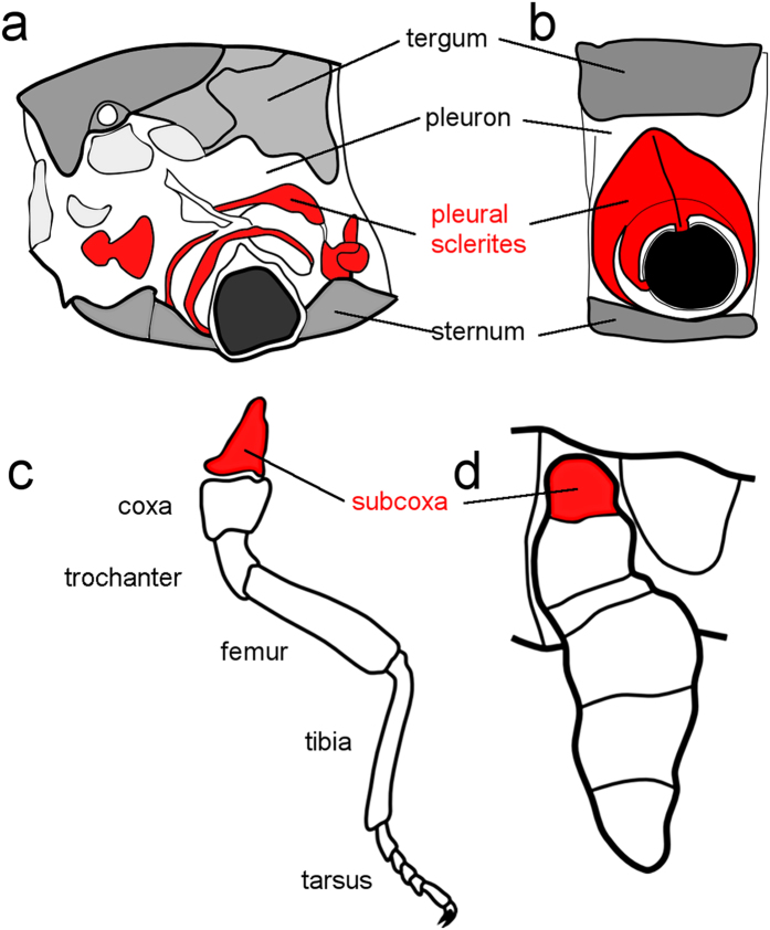
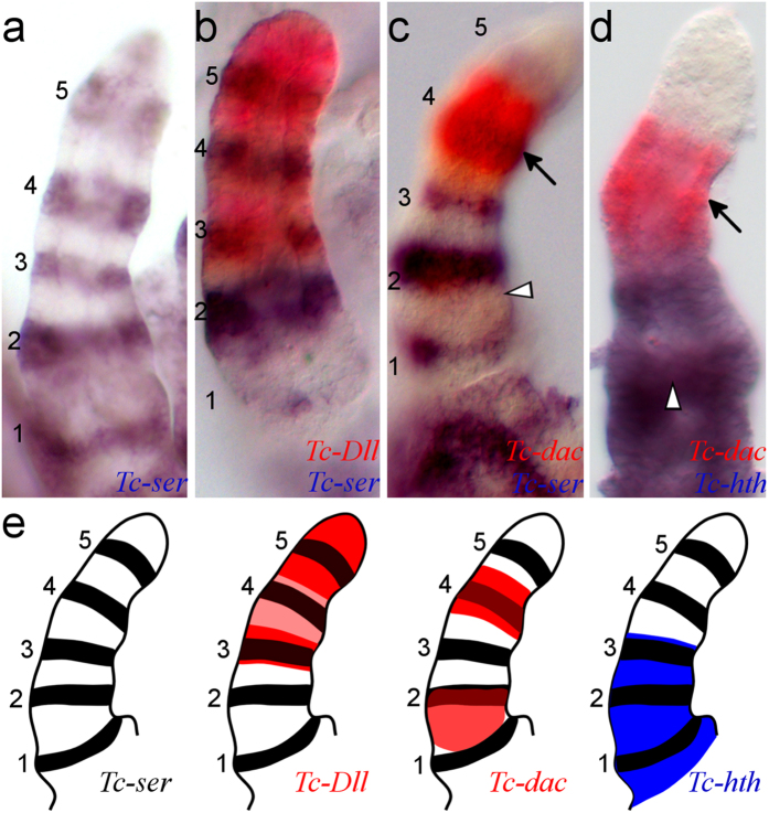
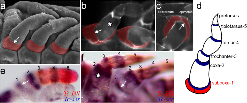
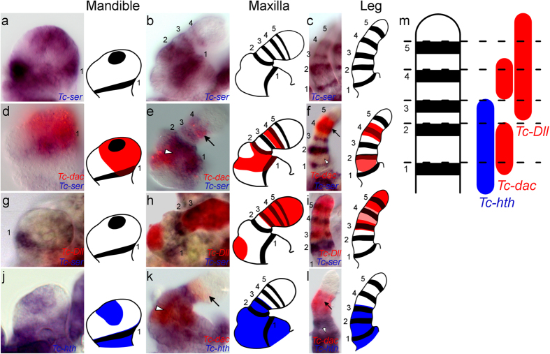
Pleurites are chitinous plates in the body wall of insects and myriapods. They are believed to be an adaptation to locomotion on land but their developmental and evolutionary origins are unclear. A widely endorsed explanation for their origin is through toughening pre-existing parts of the body wall; in contrast, the subcoxal theory suggests pleurites derive from a redeployment of the proximal-most section of the leg, the subcoxa. Here, by studying expression of appendage patterning genes in embryos and larvae of the beetle Tribolium castaneum, we provide the first molecular evidence for the existence of a cryptic subcoxal segment in developing legs. We follow this structure during development and show that the embryonic subcoxa later forms the pleurites of the larva as predicted by the subcoxal theory. Our data also demonstrate that subcoxal segments are present in all post-antennal appendages, including the first molecular evidence of a two-segmented mandible with a subcoxal segment in insects.


Figures

Figure 1
Figure 1. The subcoxal theory of the origin of the pleural sclerites.
The classic subcoxal theory states that the pleurites on the lateral insect thorax develop from an embryonic subcoxal segment. (a) Schematic showing the pleurites (red) in the mesothorax of a proturan, Eosentomon germanicum. (b) Diagram of the basic structure of a pterygote insect pleuron on a wingless thoracic segment. For clarity, the limb more distal to the subcoxa is not shown in (a,b). (c) Schematic showing segmental identities in a generalized embryonic limb. The subcoxa (which forms the pleural sclerites) is highlighted in red. (d) Schematic of the subcoxa (red) in an embryonic limb of Naucoris sp. (Rhynchota, Hemiptera). (ac) were adapted from (d) was adapted from.
Figure 2
Figure 2. Defining Tc-ser expression domains in the embryonic leg relative to limb gap gene expression.
Gene expression was detected by in situ hybridisation of dissected legs from germ band-retracted stage embryos. All views are distal to the top and lateral to the left. Domains of Tc-ser are numbered 1 to 5 from proximal to distal. The distal domain of Tc-dac is indicated with an arrow, proximal domain of Tc-dac is indicated with a white arrowhead (in (c,d)). (a) Expression of Tc-ser (blue). There are five domains of Tc-ser expression in the embryonic leg. (b) Expression of Tc-ser (blue) and Tc-Dll (red). Tc-Dll is co-expressed with the third, fourth and fifth domains of Tc-ser, but is most strongly expressed in two domains, a ‘ring’ and a ‘sock’ domain. The ring domain is co-expressed with the third Tc-ser domain, the sock domain is co-expressed with the fifth domain. (c) Expression of Tc-ser (blue) and Tc-dac (red). Tc-dac expression shows proximal and distal expression domains. The distal domain of Tc-dac is co-expressed with the fourth domain of Tc-ser whilst the proximal domain of Tc-dac expression is expressed slightly overlapping and proximal to the second Tc-ser domain (see b,d). (d) Expression of Tc-hth (blue) and Tc-dac (red). (e) Diagrams showing the gene expression of Tc-Dll, Tc-dac and Tc-hth relative to Tc-ser shown in (a,d). The distal limit of Tc-hth expression can be related to Tc-ser expression by comparison with Tc-dac expression. Tc-hth is expressed adjacent to the distal domain of Tc-dac expression. Tc-hth is therefore co-expressed with the first, second and third domains of Tc-ser.
Figure 3
Figure 3. The subcoxal segment of the embryonic leg becomes pleurites in the larva.
The white arrow indicates the boundary between the subcoxal and the coxal segments in (ac) and (e,f). A white star indicates the coxa in b and f. (a) Scanning electron micrograph (SEM) of developing leg appendages of Tribolium embryos showing the presence of a subcoxa (highlighted red) on the developing leg. Embryo at germ band retracting stage, ventral-lateral view. (b) Cuticle preparation of a first instar larva, ventral view. The pleurites are highlighted in red. (c) Cuticle preparation of a first instar larva, lateral view. The two pleural sclerites typical of ectognathous insects, the epimeron and episternum, are indicated (highlighted in red). This condition is typical of pterygote insects. (d) Schematic representation of a Tribolium larval leg with the name of the limb segments and their corresponding domains of Tc-ser expression (in blue) in the distal part of each segment. The pleurites are highlighted in red. (e) in situ hybridisation with expression of Tc-ser (blue) and Tc-Dll (red) in a dissected leg of a germ band retracted stage embryo. (f) in situ hybridisation with expression of Tc-ser (blue) and Tc-Dll (red) in a late stage embryo undergoing dorsal closure.
Figure 4
Figure 4. Serial homology of the subcoxa and coxal segment shown by leg gap gene and Tc-ser expression in the embryonic mandible, maxilla and leg.
All views are distal to the top and lateral to the right except when otherwise indicated. Domains of Tc-ser are numbered 1 to 5 from proximal to distal. The black arrow shows the distal domain of Tc-dac expression, and the white arrowhead shows the expression of the proximal domain of Tc-dac expression. Gene expression is shown schematically for each panel. Gene expression is shown for the mandible (in a,d,g and j), the maxilla (in b,e,h and k) and leg (in c,f,i and l). (a) Expression of Tc-ser (blue) in the mandible. Lateral view. (b) Expression of Tc-ser (blue) in the maxilla. (c) Expression of Tc-ser (blue) in the leg. Lateral is to the left. (d) Expression of Tc-ser (blue) and Tc-dac (red) in a dissected mandible. (e) Expression of Tc-ser (blue) and Tc-dac (red) maxilla. (f) Expression of Tc-ser (blue) and Tc-dac (red) in a leg. Lateral is to the left. (g) Expression of Tc-ser (blue) and Tc-Dll (red) in a mandible. (h) Expression of Tc-ser (blue) and Tc-Dll (red) in a maxilla. There is an endite expression domain of Tc-Dll in the second segment, which is not present in the legs. (i) Expression of Tc-ser (blue) and Tc-Dll (red) in a leg. Lateral is to the left. (j) Expression of Tc-hth (blue) and Tc-dac (red) in a dissected mandible. (k) Expression of Tc-hth (blue) and Tc-dac (red) in a maxilla. The distal limit of Tc-hth expression in the maxilla can be related to Tc-ser expression by comparison with Tc-dac expression. Tc-hth is expressed up to the trochanteral-3 domain of Tc-ser expression and therefore co-expressed with the third Tc-ser domain in the maxilla and legs. (l) Expression of Tc-hth (blue) and Tc-dac (red) in a leg. Lateral is to the left. (m) Diagram showing leg gap gene expression corresponding to domains of Tc-ser expression.

References

    1. Snodgrass R. E. Morphology and mechanism of the insect thorax. Smithsonian Miscellaneous Collect. 80, 1–108 (1927).
    1. Manton S. M. The evolution of arthropodan locomotory mechanisms. Part 9. Functional requirements and body design in Symphyla and Pauropoda and the relationships between Myriapoda and pterygote insects. J Linn Soc Lond (Zoology) 46, 103–141. (1966).
    1. Bäcker H., Fanenbruck M. & Wägele J. W. A forgotten homology supporting the monophyly of Tracheata: The subcoxa of insects and myriapods re-visited. Zool Anz 247, 185–207, 10.1016/j.jcz.2007.11.002 (2008). - DOI
    1. Manton S. M. The Arthropods: Habits, Functional Morphology and Evolution. (Clarendon, 1977).
    1. Miall L. C. & Denny A. The structure and life-history of the cockroach (Periplaneta orientalis) an introduction to the Study of Insects (Lovell Reeve and Co., 1886).

Publication types

MeSH terms

LinkOut - more resources