. 2016 Jan 1;594(1):223-37.
doi: 10.1113/JP270966. Epub 2015 Dec 14.

Deficiency of GABAergic synaptic inhibition in the Kölliker-Fuse area underlies respiratory dysrhythmia in a mouse model of Rett syndrome

Affiliations

Deficiency of GABAergic synaptic inhibition in the Kölliker-Fuse area underlies respiratory dysrhythmia in a mouse model of Rett syndrome

Ana Paula Abdala et al. J Physiol. .

Abstract

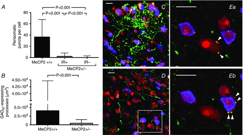
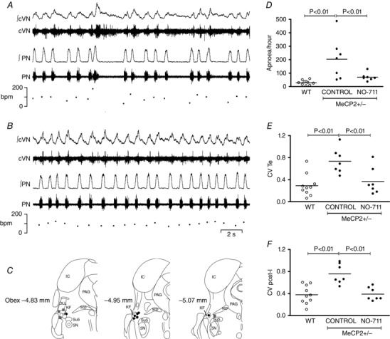
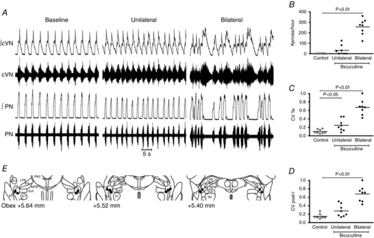
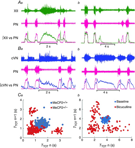
Life threatening breathing irregularity and central apnoeas are highly prevalent in children suffering from Rett syndrome. Abnormalities in inhibitory synaptic transmission have been associated with the physiopathology of this syndrome, and may underlie the respiratory disorder. In a mouse model of Rett syndrome, GABAergic terminal projections are markedly reduced in the Kölliker-Fuse nucleus (KF) in the dorsolateral pons, an important centre for control of respiratory rhythm regularity. Administration of a drug that augments endogenous GABA localized to this region of the pons reduced the incidence of apnoea and the respiratory irregularity of Rett female mice. Conversely, the respiratory disorder was recapitulated by blocking GABAergic transmission in the KF area of healthy rats. This study helps us understand the mechanism for generation of respiratory abnormality in Rett syndrome, pinpoints a brain site responsible and provides a clear anatomical target for the development of a translatable drug treatment. Central apnoeas and respiratory irregularity are a common feature in Rett syndrome (RTT), a neurodevelopmental disorder most often caused by mutations in the methyl-CpG-binding protein 2 gene (MECP2). We used a MECP2 deficient mouse model of RTT as a strategy to obtain insights into the neurobiology of the disease and into mechanisms essential for respiratory rhythmicity during normal breathing. Previously, we showed that, systemic administration of a GABA reuptake blocker in MECP2 deficient mice markedly reduced the occurrence of central apnoeas. Further, we found that, during central apnoeas, post-inspiratory drive (adductor motor) to the upper airways was enhanced in amplitude and duration in Mecp2 heterozygous female mice. Since the pontine Kölliker-Fuse area (KF) drives post-inspiration, suppresses inspiration, and can reset the respiratory oscillator phase, we hypothesized that synaptic inhibition in this area is essential for respiratory rhythm regularity. In this study, we found that: (i) Mecp2 heterozygous mice showed deficiency of GABA perisomatic bouton-like puncta and processes in the KF nucleus; (ii) blockade of GABA reuptake in the KF of RTT mice reduced breathing irregularity; (iii) conversely, blockade of GABAA receptors in the KF of healthy rats mimicked the RTT respiratory phenotype of recurrent central apnoeas and prolonged post-inspiratory activity. Our results show that reductions in synaptic inhibition within the KF induce rhythm irregularity whereas boosting GABA transmission reduces respiratory arrhythmia in a murine model of RTT. Our data suggest that manipulation of synaptic inhibition in KF may be a clinically important strategy for alleviating the life threatening respiratory disorders in RTT.


Figures

Figure 1
Figure 1. GABAergic projections in the Kölliker–Fuse nucleus (KF‐nu) of Mecp2+/−/GAD67‐eGFP compared with the same region in Mecp2+/+/GAD67‐eGFP littermate female mice
GABAergic neurones express eGFP under the control of the GAD67 promoter via a knock‐in transgene (green); perikarya are labelled in red (Nissl stain) and MECP2 protein immuno‐reactive (MECP2ir) nuclei are pseudo‐coloured in blue. This GAD67‐eGFP knock‐in strain identifies only parvalbumin GABA processes (Chattopadhyaya et al. 2004). A, quantification of the number of GABAergic bouton‐like puncta per MECP2ir positive (IR+) and negative (IR−) perikarya in the KF of Mecp2 +/−/GAD67‐eGFP versus Mecp2 +/+/GAD67‐eGFP littermate. B, quantification of the volume of GABAergic processes per field of view (4.5 × 105 μm3). C and D, representative projection of a z‐stack across the KF‐nu in a Mecp2 +/+/GAD67‐eGFP female (C) and in a Mecp2 +/−/GAD67‐eGFP littermate female (D). Reduction in MECP2ir is expected in Mecp2 +/− mice. Note the marked reduction in GFP‐expressing GABAergic projections in MECP2 deficient females. Ea and b, two sequential images of a single confocal plane of the region of interest outlined in D showing eGFP‐expressing perisomatic puncta (arrowheads) on IR+ and IR− cells. Plots show median ± interquartile range, Kruskal–Wallis test followed by Dunn's multiple comparison test (A) and Mann–Whitney test (B). Scale bars = 10 μm.
Figure 2
Figure 2. Blockade of GABA reuptake in the Kölliker–Fuse area (KF) rescued respiratory phenotype of MECP2 deficient females (Mecp2+/–, n = 7) to wild‐type (WT, n = 10) levels
A, typical trace showing resting phrenic nerve (PN), central vagus nerve (cVN) and respiratory rate in a Mecp2 +/− female mouse. B, the same Mecp2 +/– mouse after microinjection of NO‐711 (GABA reuptake blocker, 10 μm, 60 nL) in the KF. C, schematic diagram modified from (Franklin & Paxinos, 2007) showing effective (black circles) and non‐effective (grey squares) microinjection sites (section distances from obex). Abbreviations: 5 N, motor trigeminal nucleus; DLL, dorsal nucleus of the lateral lemniscus; IC, inferior colliculus; PAG, periaqueductal grey; scp, superior cerebellar peduncle; Su5, supratrigeminal nucleus. D, blockade of GABA reuptake in the KF reduced occurrence of apnoeas in Mecp2 +/– female mice (filled circles, n = 7) to the level of littermate wild‐type females (open circles, n = 10). E, microinjections also reduced the coefficient of variation (CV) of expiratory time (T E) in the PN, and the CV of post‐inspiration (post‐I) in the cVN to that of WT females (F). Plots show individual values (circles) and means (lines), one‐way ANOVA, Newman–Keuls all pairwise comparison.
Figure 3
Figure 3. Blockade of GABAA receptors in the Kölliker–Fuse area (KF) induced Rett‐like respiratory phenotype in wild‐type rats ( n = 8)
A, typical trace showing phrenic nerve (PN), central vagus nerve (cVN) during a control period and after unilateral and bilateral microinjection of bicuculline (5 mm, 60 nL), a GABAA receptor antagonist. B, central apnoeas were absent in wild‐type rats (open circles), and unilateral and bilateral blockade of GABAA receptors induced central apnoeas in 4/8 and 8/8 rats, respectively (filled circles). C and D, microinjections also increased the coefficient of variation of expiratory time (T E) in the PN (C), and the coefficient of variation of post‐inspiration (post‐I) (D). E, schematic diagram modified from Franklin and Paxinos (2007) showing effective (black circles) and non‐effective (grey squares) bilateral microinjection sites. Abbreviations: 5 N, motor trigeminal nucleus; A7, facial motor nucleus; DLL, dorsal nucleus of the lateral lemniscus; MPB, medial parabrachial nucleus; LPB, lateral parabrachial nucleus; PAG, periaqueductal grey; scp, superior cerebellar peduncle; Su5, supratrigeminal nucleus. Plots show individual values (circles) and means (lines), one‐way ANOVA, Dunnett's pairwise comparison versus control.
Figure 4
Figure 4. Example histological photomicrographs of microinjection sites in the Kölliker–Fuse area (KF)
A and B, dorsolateral pons of a Mecp2 +/– female mouse counterstained with DAPI (A) showing microinjection site labelled with red fluorescent beads (x) (B). C and D, dorsolateral pons of a Wistar rat counterstained with DAPI (C) showing microinjection site marked with red fluorescent beads (x) (D). Anatomical landmarks: ll, lateral lemniscus; me5, mesencephalic trigeminal nucleus; MPB, medial parabrachial nucleus; LPB, lateral parabrachial nucleus; scp, superior cerebellar peduncle. Scale bars = 500 μm.
Figure 5
Figure 5. Comparison of respiratory motor outputs in Mecp2+/− mice versus effects of blocking Kölliker–Fuse area (KF) GABAA receptors in wild‐type (WT) rats
A, typical traces of the hypoglossal (XII) and phrenic (PN) nerve activities during an apnoea in a Mecp2 +/− mouse (Aa) and after bilateral blockade of KF GABAA receptors (bicuculline, 5 mm, 60 nL) in a WT rat (Ab). Note XII activity during apnoea, and temporary loss inspiratory drive in the first breath after apnoea. B, typical traces of the cervical vagus (cVN) and phrenic (PN) nerve activities during an apnoea in a Mecp2 +/− mouse (Ba) and after bilateral blockade of KF GABAA receptors in a WT rat (Bb). Note post‐inspiratory activity in the cVN throughout the apnoeas. C, Poincaré plots of total respiratory period (T TOT) in a Mecp2 +/− mouse (Ca) presenting periodic breathing, and after bilateral blockade of KF GABAA receptors in a WT rat (Cb). Note similar scattering of PN period in Mecp2 +/− and in bicuculline induced breathing dysrhythmia in rats, typical of periodic breathing.

References

    1. Abdala AP, Bissonnette JM & Newman‐Tancredi A (2014. a). Pinpointing brainstem mechanisms responsible for autonomic dysfunction in Rett syndrome: therapeutic perspectives for 5‐HT1A agonists. Front Physiol 5, 205. - PMC - PubMed
    1. Abdala AP, Dutschmann M, Bissonnette JM & Paton JF (2010). Correction of respiratory disorders in a mouse model of Rett syndrome. Proc Natl Acad Sci USA 107, 18208–18213. - PMC - PubMed
    1. Abdala AP, Lioy DT, Garg SK, Knopp SJ, Paton JF & Bissonnette JM (2014. b). Effect of Sarizotan, a 5‐HT1a and D2‐like receptor agonist, on respiration in three mouse models of Rett syndrome. Am J Respir Cell Mol Biol 50, 1031–1039. - PMC - PubMed
    1. Alheid GF, Gray PA, Jiang MC, Feldman JL & McCrimmon DR (2002). Parvalbumin in respiratory neurons of the ventrolateral medulla of the adult rat. J Neurocytol 31, 693–717. - PubMed
    1. Ballas N, Lioy DT, Grunseich C & Mandel G (2009). Non‐cell autonomous influence of MeCP2‐deficient glia on neuronal dendritic morphology. Nat Neurosci 12, 311–317. - PMC - PubMed