. 2015 Nov 10;112(45):14078-83.
doi: 10.1073/pnas.1512812112. Epub 2015 Oct 27.

Allosteric activation of M4 muscarinic receptors improve behavioral and physiological alterations in early symptomatic YAC128 mice

Affiliations

Allosteric activation of M4 muscarinic receptors improve behavioral and physiological alterations in early symptomatic YAC128 mice

Tristano Pancani et al. Proc Natl Acad Sci U S A. .

Abstract

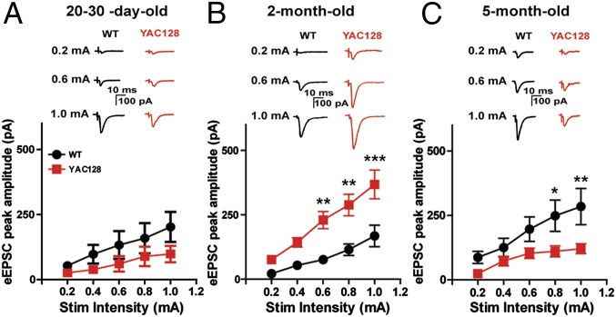
Mutations that lead to Huntington's disease (HD) result in increased transmission at glutamatergic corticostriatal synapses at early presymptomatic stages that have been postulated to set the stage for pathological changes and symptoms that are observed at later ages. Based on this, pharmacological interventions that reverse excessive corticostriatal transmission may provide a novel approach for reducing early physiological changes and motor symptoms observed in HD. We report that activation of the M4 subtype of muscarinic acetylcholine receptor reduces transmission at corticostriatal synapses and that this effect is dramatically enhanced in presymptomatic YAC128 HD and BACHD relative to wild-type mice. Furthermore, chronic administration of a novel highly selective M4 positive allosteric modulator (PAM) beginning at presymptomatic ages improves motor and synaptic deficits in 5-mo-old YAC128 mice. These data raise the exciting possibility that selective M4 PAMs could provide a therapeutic strategy for the treatment of HD.

Keywords: basal ganglia; movement disorder; neurodegenerative; trinucleotide repeat disorder.


Conflict of interest statement

Conflict of interest statement: P.J.C. has been funded by the NIH, Johnson & Johnson, AstraZeneca, Bristol-Myers Squibb, Michael J. Fox Foundation, and Seaside Therapeutics. Over the past 3 years, he has served on the Scientific Advisory Boards of Seaside Therapeutics, Michael J. Fox Foundation, Stanley Center for Psychiatric Research at Broad Institute (MIT/Harvard), Karuna Pharmaceuticals, Lieber Institute for Brain Development (Johns Hopkins University), Clinical Mechanism and Proof of Concept Consortium, and Neurobiology Foundation for Schizophrenia and Bipolar Disorder. He is an inventor on patents that protect different classes of muscarinic receptor allosteric modulators.

Figures

Fig. 1.
Fig. 1.
Age-dependent biphasic alteration in corticostriatal glutamatergic transmission. (Top) Representative traces of EPSCs evoked with increasing stimulus intensities (0.2, 0.6, and 1.0 mA) in dorsal SPNs from 20- to 30-d-old (A), 2-mo-old (B), and 5-mo-old (C) WT (black) and YAC128 (red) mice. (Bottom) Input–output graphs showing increased eEPSC amplitude in SPNs from 2-mo-old YAC128 (B) and a decrease in 5-mo-old YAC128 compared with WT (C). No alterations are seen in 20- to 30-d-old animals (A) (*P < 0.05, **P < 0.01, ***P < 0.001, Bonferroni posttest). Data are reported as mean ± SEM.
Fig. S1.
Fig. S1.
Increased corticostriatal glutamatergic transmission in 2-mo-old BACHD mice is accompanied by an increase in M4-mediated modulation. (A) Representative traces of EPSCs evoked with increasing stimulation intensities (0.2, 0.6, and 1 mA) in dorsal SPNs from 2-mo-old WT (black) and BACHD (red) mice. (B) I/O graphs showing increased eEPSC peak amplitude in 2-mo-old BACHD (red; n = 12) compared with WT (black; n = 10) [main effect of genotype two-way RM ANOVA, F(1,100) = 35.59, P < 0.0001; *P < 0.05, ***P < 0.001, ****P < 0.0001, Bonferroni posttest]. In gray, the I/O graph shows increased eEPSC peak amplitude in YAC128FVB (n = 4) compared with WT (n = 10) [main effect of genotype in a two-way RM ANOVA, F(1,60) = 23.50, P < 0.0001; Bonferroni posttest reports P < 0.05 at 0.8 mA and P < 0.01 at 1 mA]. (C) Representative traces showing the maximal inhibitory effect of 3 μM CCh alone or in combination with 3 μM VU0467154 (VU154) on eEPSC peak amplitude in WT (black) and 2-mo-old BACHD (red) mice. (D) Graph showing the potentiating effect of VU0467154 on CCh-mediated inhibition of peak eEPSCs in SPNs from BACHD mice (*P < 0.05 compared with CCh alone, t test) but not in neurons from WT mice (P > 0.05 compared with CCh alone, t test). Similarly, the maximal inhibitory effect of 3 μM CCh on eEPSC peak amplitude was greater in BACHD mice compared with WT (#P < 0.05, t test). In gray is shown the inhibitory effect of 3 μM CCh on eEPSCs in SPNs from YAC128FVB mice (#P < 0.05 compared with WT, t test). n = 4–8 in C and D. Data are reported as mean ± SEM.
Fig. S2.
Fig. S2.
sEPSC amplitude changes. (Left) Representative traces showing sEPSCs in SPNs. (Right) Box-and-whisker graphs summarizing the increase in sEPSC amplitude in SPNs from 2-mo-old YAC128 compared with age-matched WT (B). No changes were noted at other ages (A and C) (#P = 0.001, Mann–Whitney nonparametric test). (B, Bottom) No change in sEPSC frequency in SPNs from 2-mo-old YAC128 mice (P > 0.05, Mann–Whitney nonparametric test). Data are reported as mean ± SEM.
Fig. S3.
Fig. S3.
Somatic afterhyperpolarization (AHP) is decreased in YAC128 mice at 2 mo of age. (Left) Representative traces of AHP currents from slices from WT (black) and YAC128 (red) mice. (Right) Bar graph summarizing mean peak amplitude of somatic AHP currents in SPNs from 2-mo-old WT and YAC128 mice. AHP current was elicited with a 10-ms voltage step to +10 mV from a holding potential of −70 mV in voltage-clamp configuration (*P < 0.05, t test). (Right) Bar graph summarizing mean peak amplitude of somatic AHP current in SPNs from 5-mo-old WT and YAC128 mice (WT, n = 9; YAC, n = 10; P > 0.05, t test). Data are reported as mean ± SEM.
Fig. S4.
Fig. S4.
Positive correlation between corticostriatal glutamate and rotarod performance. (A) Bar graph summarizing the impairment in motor coordination evidenced by a decrease in time to fall (TTF) on an accelerated rotarod in 5-mo-old YAC128 (red) mice compared with age-matched WT (black) (**P < 0.01, t test). (B) Representative graph showing a comparison of eEPSC I/O curves obtained by averaging the I/O from each SPN recorded from WT mouse ID CY576N and YAC128 mouse ID CY612N. The WT mouse showed greater eEPSC amplitudes overall and higher TTF (102 s), whereas the YAC128 mouse showed decreased eEPSC amplitudes and lower TTF (56 s). (C) Graph showing a positive correlation between rotarod TTF and eEPSC peak amplitude at the corticostriatal synapse (R2 = 0.47, P = 0.03) in SPNs from WT (black) and YAC128 (red) mice. TTF was ranked from worst-performing (low rank, shorter TTF) to best-performing (high rank, longer TTF), whereas glutamatergic transmission was ranked as follows: The total glutamatergic transmission obtained from each neuron by averaging peak EPSC amplitudes across each stimulus in the I/O curve was averaged with other cells within each animal and ranked. Data are reported as mean ± SEM.
Fig. 2.
Fig. 2.
Age-dependent increase in M4-mediated modulation of corticostriatal glutamatergic transmission. (A, Left) Representative traces showing the inhibitory effect of 3 μM CCh on eEPSC peak amplitude in SPNs from WT (black) and YAC128 (red) in 20- to 30-d-old and 2-mo-old mice. (A, Right) Graph summarizing the effects of 3 μM CCh on eEPSCs recorded in SPNs from 20- to 30-d-old and 2-mo-old mice (*P < 0.05, paired t test). (B) Bar graph summarizing the potentiating effect of 3 μM VU0467154 on the CCh (1 μM)-mediated inhibition of eEPSC amplitude in WT and YAC128 mice in SPNs from WT and YAC128 mice (**P < 0.01, t test). (C) Representative traces showing the inhibitory effect of CCh alone or in combination with 3 μM VU0467154 (VU154) on eEPSC peak amplitude. (D) Graph showing the effect of 1 μM CCh in combination with 3 μM VU0467154 on the paired-pulse ratio in SPNs from WT (black) and YAC128 (red) (*P < 0.05, paired t test). Data are reported as mean ± SEM.
Fig. S5.
Fig. S5.
CCh-mediated inhibition of eEPSC amplitude in slices from 2- and 5-mo-old mice. (A) Inhibitory effect of increasing concentration of CCh (1–10 μM) on corticostriatal eEPSCs in SPNs from 2-mo-old WT (black) and YAC128 (red) (*P < 0.05, **P < 0.01, t test). (B) Inhibitory effect of CCh (3 μM) on corticostriatal eEPSCs in SPNs from 5-mo-old WT and YAC128 animals; no significant difference was seen (P > 0.05, t test). Data are reported as mean ± SEM.
Fig. 3.
Fig. 3.
Corticostriatal glutamatergic transmission is increased in 2-mo-old YAC128/M4/ mice. (A) Representative eEPSC traces elicited with 1-mA stimulation intensity in SPNs from YAC128 (red) and YAC128/M4/ (blue), as well as in WT (gray) shown for comparison. (B) I/O graph showing increased eEPSCs elicited with increasing stimulation intensities (0.2–1 mA) in SPNs from YAC128/M4/ compared with YAC128 mice (*P < 0.05, **P < 0.01, Bonferroni posttest). WT I/O curve is shown for comparison. (C) No effect of 3 μM CCh on peak eEPSC amplitude was seen in SPNs from YAC128/M4/. (Insets) Representative traces recorded from baseline and during CCh application. Data are reported as mean ± SEM.
Fig. S6.
Fig. S6.
Chronic administration of VU0467154 ameliorates deficits in motor coordination and locomotor activity in 5-mo-old YAC128 mice. (A) Experimental paradigm. Accelerated rotarod with constant increase in revolutions per min. (B, Left) Bar graph summarizing average TTF in the accelerated rotarod test in the first trial on test day in 2-mo-old WT and YAC128 mice. No significant alterations were noted in YAC128 compared with WT mice (P > 0.05, t test). (B, Right) Bar graph summarizing total distance traveled in 2-mo-old WT and YAC128 mice. No alterations were noted in YAC128 compared with WT (P > 0.05, t test). (C) Time course of the open-field experiments in 5-mo-old mice displaying the average distance traveled per treatment group across the course of the entire 60-min experiment. (D) Bar graph summarizing rearing counts across all four treatment groups. Chronic treatment with VU0467154 was able to normalize rearing behavior in 5-mo-old YAC128 but did not have any effect in WT mice (*P < 0.05, Bonferroni posttest). Data are reported as mean ± SEM.
Fig. 4.
Fig. 4.
Chronic VU0467154 improves striatal glutamatergic and dopaminergic signaling in early symptomatic YAC128 mice. (A) Representative traces showing eEPSCs evoked with 1-mA stimulus intensity recorded from dorsal SPNs from mice chronically treated with either VU0467154 (VU154) or vehicle. (B) I/O graph showing decreased corticostriatal glutamatergic transmission in YAC128 mice chronically treated with VU0467154 (YAC128-VU154; *P < 0.05, Bonferroni posttest). (C and D) Graphs showing DA release (overflow) evoked with increasing stimulus intensities (25–800 μA) in Vh-treated (C) and VU154-treated (D) WT and YAC128 mice. Vh-treated YAC128 mice display a consistent decrease in DA release across all stimulation intensities, with a significant decrease observed at 800-μA stimulation intensity (*P < 0.05, Bonferroni posttest) compared with Vh-treated WT. No statistical difference was seen in VU154-treated WT or YAC128 mice (D). (Insets) Representative traces showing an electrically evoked (800-μA stimulation; arrows) DA rise in slices from Vh-treated WT (black) and YAC128 mice (red) and from VU0467154-treated WT and YAC128 mice. Data are reported as mean ± SEM.
Fig. S7.
Fig. S7.
Chronic treatment with VU0467154 does not alter muscarinic modulation of glutamate transmission in either WT or YAC128 mice. The inhibitory effect of 3 μM CCh on eEPSC peak amplitude in SPNs recorded in Vh- and VU0467154-treated 5-mo-old WT and YAC128 mice is not significantly different across treatment or genotype (two-way ANOVA). Data are reported as mean ± SEM.
Fig. 5.
Fig. 5.
Chronic administration of VU0467154 improves motor coordination and locomotor activity in 5-mo-old YAC128 mice. (A) Vh-treated YAC128 mice displayed reduced time spent on the rotarod compared with Vh-treated WT animals (#P < 0.05, t test). Chronic treatment with VU0467154 significantly increased the time spent on the rotarod in YAC128 but not in WT mice (*P < 0.05, Bonferroni posttest). (B) Five-month-old Vh-treated YAC128 animals displayed reduced total distance traveled (#P < 0.05, t test) compared with Vh-treated WT mice. Chronic treatment with VU0467154 (10 mg/kg) improved locomotor activity in YAC128 but not in WT animals (*P < 0.05, Bonferroni posttest). (C) Representative locomotor activity paths from one representative 5-mo-old animal per treatment group. Dots represent the position of the mouse at the end of the experiment. Data are reported as mean ± SEM.
Fig. S8.
Fig. S8.
Acute administration of VU0467154 does not affect accelerated rotarod performance in mice. No significant effect of 10 mg/kg VU0467154 i.p. was seen 45 min after injection (a time sufficient to reach peak levels of VU0467154 in the CNS) in a small cohort of symptomatic YAC128 mice (n = 4 per treatment group, P > 0.05, t test). Vh-treated WT mouse performance is shown for comparison (n = 5). Although the number of mice was limited, a significant impairment in rotarod performance (decreased time to fall) was detected in Vh-treated YAC128 compared with WT (P = 0.02, t test). Data are reported as mean ± SEM.
Fig. S9.
Fig. S9.
Photomicrographs of immunofluorescence staining representing images of striatal parenchyma showing no change in Fluoro-Jade C and NeuN fluorescence across all treatment groups in mice chronically treated with Vh or VU0467154. (Bottom) A dramatic increase in Fluoro-Jade C–positive cells along with decreased NeuN-positive cells in the striatum from an 8-wk-old R6/2 mouse is shown for comparison.
Fig. S10.
Fig. S10.
Graphs showing quantification of NeuN- and Fluoro-Jade C–positive cells per field of view as well as quantification of mHtt inclusion-positive cells per field of view in the striatum from two Vh-treated WT, two Vh-treated YAC128, and two VU0467154-treated WT and YAC128. There was no significant change in NeuN-positive neurons across all treatment groups (one-way ANOVA with a Tukey’s post hoc test). Moreover, no detectable Fluoro-Jade C–positive cells were seen in YAC128 and WT mice as well as no nuclear mHtt inclusions, and no effect of VU0467154 treatment (one-way ANOVA with a Tukey’s post hoc test). As a positive control, we evaluated the striatum from an 8-wk-old R6/2 mouse and found a decrease in NeuN-positive cells (see also Fig. S8), the presence of intense cellular Fluoro-Jade C staining in the striatum, and increased mHtt nuclear deposits. This is consistent with previous studies showing the presence of these pathological markers in R6/2 mice. Altogether, these data show that in 5-mo-old YAC128 significant striatal pathology is absent. Data are reported as mean ± SEM.
Fig. S11.
Fig. S11.
MAB5374 immunoreactivity staining. Representative images of striatal parenchyma showing increased mHtt staining in YAC128 (Vh- or VU0467154-treated; C and D, compared to A and B respectively). No change in mHtt staining is present after chronic treatment with VU0467154 (D) compared with Vh-treated (C). YAC128 mice show no MAB5374-positive nuclear mHtt inclusions (F), which are clearly present in the striatum from an 8-wk-old R6/2 mouse also displaying more intense mHtt staining (E and G). The increase in mHtt in YAC128 could play a role in pathophysiological alterations in synaptic and motor function seen at this age.

References

    1. The Huntington’s Disease Collaborative Research Group A novel gene containing a trinucleotide repeat that is expanded and unstable on Huntington’s disease chromosomes. Cell. 1993;72(6):971–983. - PubMed
    1. Beglinger LJ, et al. Huntington Study Group Investigators Earliest functional declines in Huntington disease. Psychiatry Res. 2010;178(2):414–418. - PMC - PubMed
    1. Siemers E, et al. Motor changes in presymptomatic Huntington disease gene carriers. Arch Neurol. 1996;53(6):487–492. - PubMed
    1. Kirkwood SC, et al. Subtle changes among presymptomatic carriers of the Huntington’s disease gene. J Neurol Neurosurg Psychiatry. 2000;69(6):773–779. - PMC - PubMed
    1. Walker FO. Huntington’s disease. Semin Neurol. 2007;27(2):143–150. - PubMed

Publication types

Substances