Review
. 2016;51(1):26-42.
doi: 10.3109/10409238.2015.1102859. Epub 2015 Oct 28.

Sites of retroviral DNA integration: From basic research to clinical applications

Affiliations
Review

Sites of retroviral DNA integration: From basic research to clinical applications

Erik Serrao et al. Crit Rev Biochem Mol Biol. 2016.

Abstract

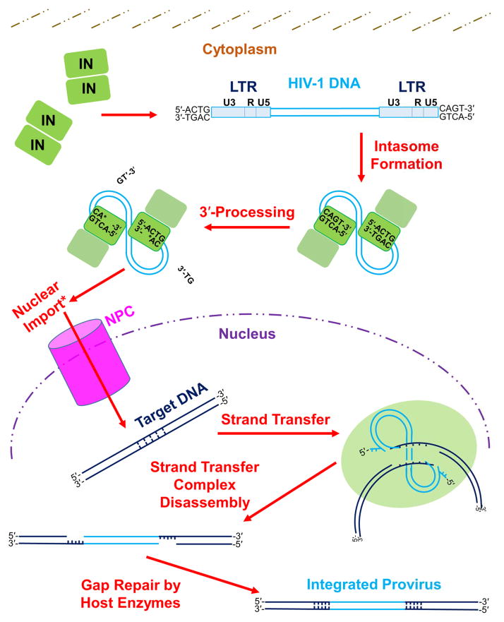
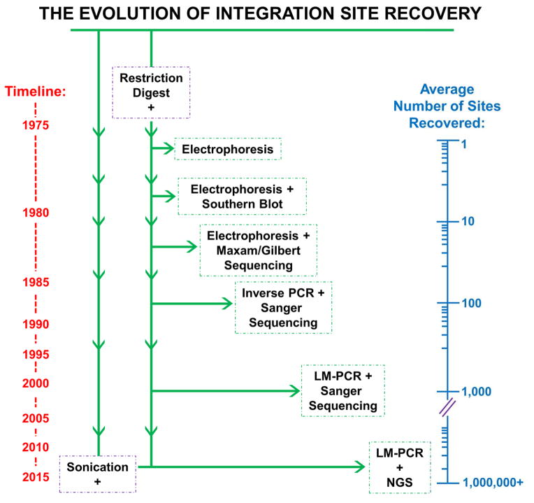
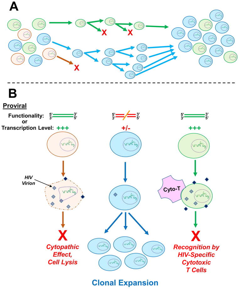
One of the most crucial steps in the life cycle of a retrovirus is the integration of the viral DNA (vDNA) copy of the RNA genome into the genome of an infected host cell. Integration provides for efficient viral gene expression as well as for the segregation of viral genomes to daughter cells upon cell division. Some integrated viruses are not well expressed, and cells latently infected with human immunodeficiency virus type 1 (HIV-1) can resist the action of potent antiretroviral drugs and remain dormant for decades. Intensive research has been dedicated to understanding the catalytic mechanism of integration, as well as the viral and cellular determinants that influence integration site distribution throughout the host genome. In this review, we summarize the evolution of techniques that have been used to recover and map retroviral integration sites, from the early days that first indicated that integration could occur in multiple cellular DNA locations, to current technologies that map upwards of millions of unique integration sites from single in vitro integration reactions or cell culture infections. We further review important insights gained from the use of such mapping techniques, including the monitoring of cell clonal expansion in patients treated with retrovirus-based gene therapy vectors, or patients with acquired immune deficiency syndrome (AIDS) on suppressive antiretroviral therapy (ART). These insights span from integrase (IN) enzyme sequence preferences within target DNA (tDNA) at the sites of integration, to the roles of host cellular proteins in mediating global integration distribution, to the potential relationship between genomic location of vDNA integration site and retroviral latency.

Keywords: Gene therapy; HIV-1; illumina; intasome; integrase; integration sites; next-generation sequencing; retrovirus.


Conflict of interest statement

Declaration of interest

The authors declare no conflict of financial interest. This work was supported by US National Institutes of Health grants AI052014 and AI039394 (to A.N.E.) as well as AI007386 (to E.S.).

Figures

Figure 1
Figure 1. The mechanism of retroviral integration
The integration process begins in the host cell with formation of the intasome, which consists of multimerized IN bound to the viral LTR ends. The LTR is composed of U3, R, and U5 sections, each of which contains unique elements responsible for mediating viral gene transcription (Pereira et al., 2000). While within the cytoplasm, IN cleaves nucleotides (specifically two nucleotides for the depicted HIV-1 integration pathway) from each 3′ end of the vDNA adjacent to invariant 5′-CA-3′ dinucleotides during 3′-processing. Completion of 3′-processing leaves a recessed and chemically reactive hydroxyl group at each vDNA 3′ end. Lentiviral PICs are actively imported into the cell nucleus (denoted by an asterisk in figure), while γ-retroviruses can only enter the nucleus after dissolution of the nuclear membrane during cell division [reviewed in: (Matreyek and Engelman, 2013)]. Within the nucleus, IN binds tDNA and utilizes the reactive vDNA 3′-hydroxyl groups to simultaneously cleave the tDNA phosphodiester backbone and insert the vDNA molecule, through the process of strand transfer. IN subunits cleave the top and bottom tDNA strands in a staggered fashion. The length of stagger differs among retroviruses; HIV-1 IN cleaves tDNA with the depicted 5-bp stagger. After disassembly of the strand transfer complex, host cell enzymes repair the DNA recombination intermediate to yield the integrated provirus flanked by a host DNA TSD. See main text for descriptions of the roles that other cellular factors play in the process of retroviral DNA integration. Please note that a color version of Figure 1 is available online.
Figure 2
Figure 2. The evolution of techniques used to determine retroviral integration sites
Integration site recovery techniques are listed in chronological order from top to bottom, corresponding to a general timeline shown at the left of the figure. The average numbers of integration sites recovered in studies employing these techniques, which exponentially increased over time, are illustrated on a logarithmic axis at the right side of the figure. The initial step of host cell gDNA fragmentation by restriction endonuclease digestion is shared by all of the listed techniques, though sonication has recently become a desirable method of shearing. Please note that a color version of this figure is available online.
Figure 3
Figure 3. General integration site preferences of different retroviral genera
Representational integration site patterns with respect to the structure of the average host cell gene are depicted by vertical arrows, with each arrow type corresponding to a different viral genus (or group of genera in the case of α-, β-, and δ-retroviruses). Gammaretroviruses preferentially target active promoters and enhancers (Wu et al., 2003, LaFave et al., 2014, De Ravin et al., 2014) while lentiviruses integrate primarily within the bodies of actively transcribed genes (Schroder et al., 2002). Spumaviruses avoid genes and rather target intergenic regions, while α-, β-, and δ-retroviruses exhibit nearly random distributions. As mentioned in the text, additional features not depicted in the figure can also influence the integration sites of various viruses, such as gene transcription level, proximity to the NPC, and local nucleotide sequence.
Figure 4
Figure 4. Clonal expansion of provirus-harboring cells
A) General schematic of cellular clonal expansion. From an initially diverse population of cells, certain clones can exhibit relatively increased viability or growth rate, thus leading to persistence and ultimate enrichment. In this example the orange cell clone is dividing slowly or not at all, while the green cell clone exhibits an intermediate rate of expansion, and the blue cell clone expands most quickly. Thus in the final cell population, the blue clone predominates. B) Host cells harboring a particular integration site(s) can also become enriched over time within infected patients. The fact that clonally expanded, provirus-harboring cells can persist in the face of suppressive ART suggests that additional factors beyond integration site-mediated stimulation of cell growth rate contribute to clonal persistence. Two suggestions have been that the resident proviruses within the expanded cell clones are 1) nonfunctional due to non-tolerable insertions or deletions or 2) are molecularly intact but are expressed at a relatively low level (or not at all). In either case, the clones would escape viral replication-associated cytopathic effects and cell lysis (orange clone), as well as host cell recognition and purging by HIV-specific cytotoxic T-cells (green clone), leading to the enrichment of particular integration sites over time (blue clone). Neither of these possibilities, though, has thus far been definitively proven. A color version of this figure is available online.

References

    1. AIUTI A, BIASCO L, SCARAMUZZA S, FERRUA F, CICALESE MP, BARICORDI C, DIONISIO F, CALABRIA A, GIANNELLI S, CASTIELLO MC, BOSTICARDO M, EVANGELIO C, ASSANELLI A, CASIRAGHI M, DI NUNZIO S, CALLEGARO L, BENATI C, RIZZARDI P, PELLIN D, DI SERIO C, SCHMIDT M, VON KALLE C, GARDNER J, MEHTA N, NEDUVA V, DOW DJ, GALY A, MINIERO R, FINOCCHI A, METIN A, BANERJEE PP, ORANGE JS, GALIMBERTI S, VALSECCHI MG, BIFFI A, MONTINI E, VILLA A, CICERI F, RONCAROLO MG, NALDINI L. Lentiviral hematopoietic stem cell gene therapy in patients with Wiskott-Aldrich syndrome. Science. 2013;341:1233151. - PMC - PubMed
    1. AIYER S, ROSSI P, MALANI N, SCHNEIDER WM, CHANDAR A, BUSHMAN FD, MONTELIONE GT, ROTH MJ. Structural and sequencing analysis of local target DNA recognition by MLV integrase. Nucleic Acids Res. 2015;43:5647–5663. - PMC - PubMed
    1. AIYER S, SWAPNA GVT, MALANI N, ARAMINI JM, SCHNEIDER WM, PLUMB MR, GHANEM M, LARUE RC, SHARMA A, STUDAMIRE B, KVARATSKHELIA M, BUSHMAN FD, MONTELIONE GT, ROTH MJ. Altering murine leukemia virus integration through disruption of the integrase and BET protein family interaction. Nucleic Acids Res. 2014;42:5917–5928. - PMC - PubMed
    1. AKAGI K, SUZUKI T, STEPHENS RM, JENKINS NA, COPELAND NG. RTCGD: retroviral tagged cancer gene database. Nucleic Acids Res. 2004;32:D523–D527. - PMC - PubMed
    1. ALBERS J, DANZER C, RECHSTEINER M, LEHMANN H, BRANDT LP, HEJHAL T, CATALANO A, BUSENHART P, GONÇALVES AF, BRANDT S, BODE PK, BODE-LESNIEWSKA B, WILD PJ, FREW IJ. A versatile modular vector system for rapid combinatorial mammalian genetics. J Clin Invest. 2015;125:1603–1619. - PMC - PubMed

Publication types

LinkOut - more resources