. 2015 Dec 8;132(23):2237-47.
doi: 10.1161/CIRCULATIONAHA.115.016540. Epub 2015 Oct 27.

Genetic Ablation of Extra Domain A of Fibronectin in Hypercholesterolemic Mice Improves Stroke Outcome by Reducing Thrombo-Inflammation

Affiliations

Genetic Ablation of Extra Domain A of Fibronectin in Hypercholesterolemic Mice Improves Stroke Outcome by Reducing Thrombo-Inflammation

Nirav Dhanesha et al. Circulation. .

Abstract

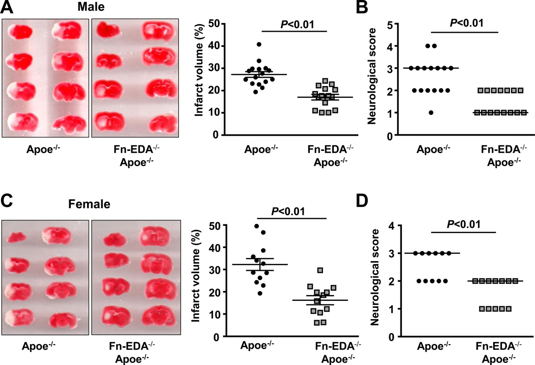
Background: The fibronectin-splicing variant containing extra domain A (Fn-EDA) is present in negligible amounts in the plasma of healthy humans but markedly elevated in patients with comorbid conditions, including diabetes mellitus and hypercholesterolemia, which are risk factors for stroke. It remains unknown, however, whether Fn-EDA worsens stroke outcomes in such conditions. We determined the role of Fn-EDA in stroke outcome in a model of hypercholesterolemia, the apolipoprotein E-deficient (Apoe(-/-)) mouse.

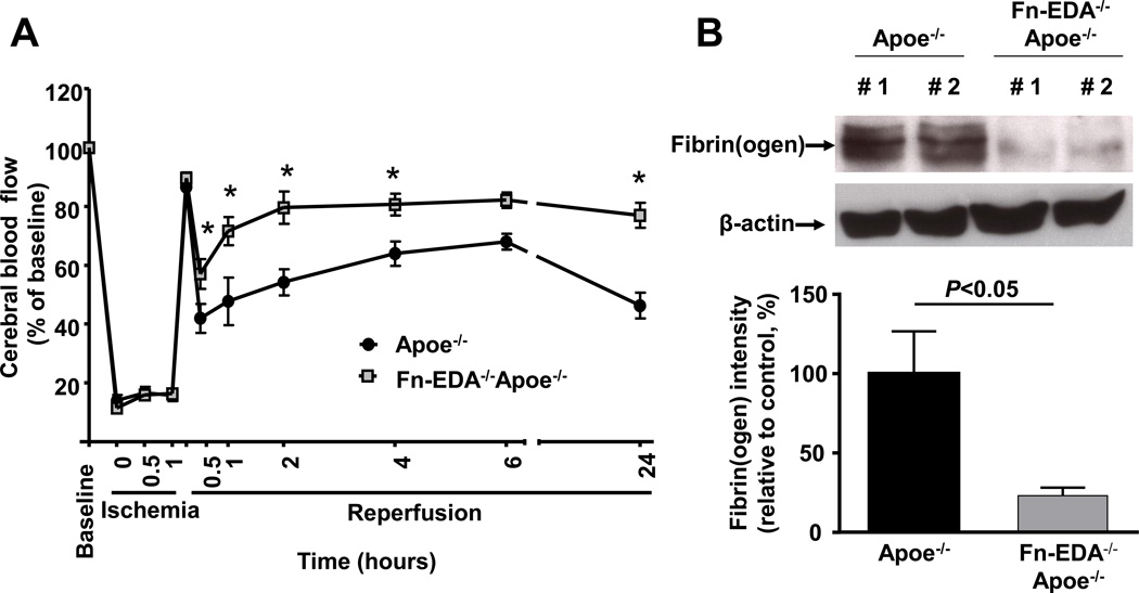
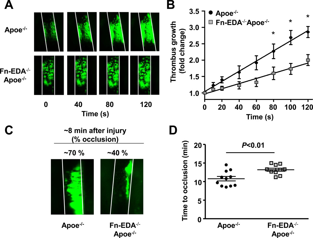
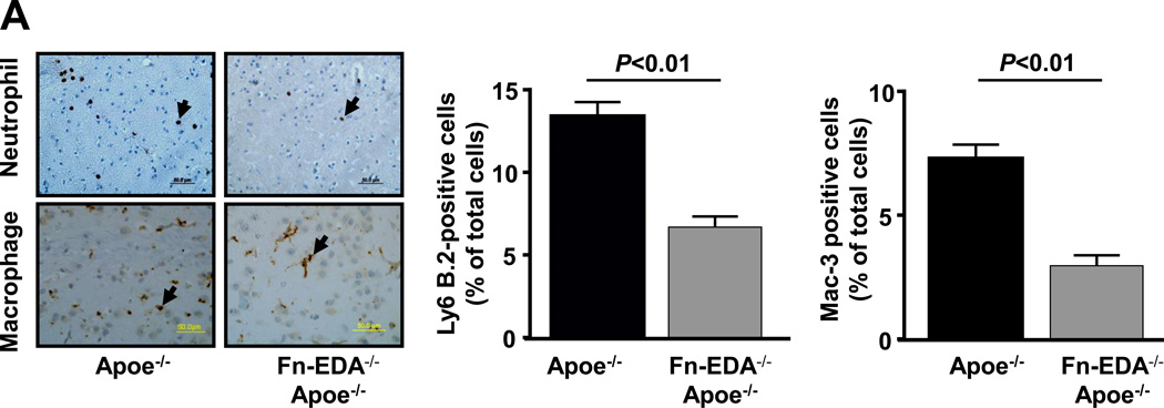
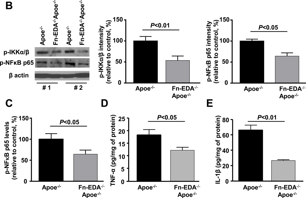
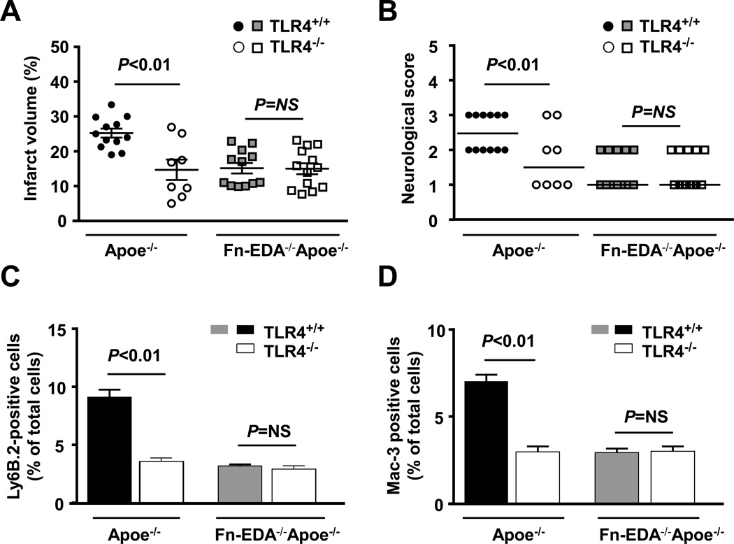
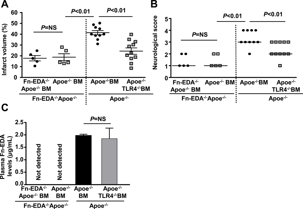
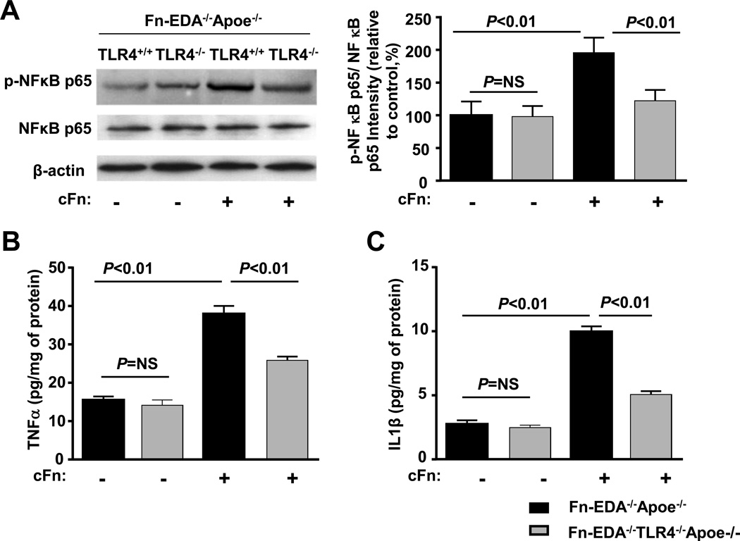
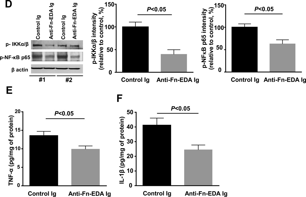
Methods and results: In a transient cerebral ischemia/reperfusion injury model, Apoe(-/-) mice expressing fibronectin deficient in EDA (Fn-EDA(-/-)Apoe(-/-) mice) exhibited smaller infarcts and improved neurological outcomes at days 1 and 8 (P<0.05 versus Apoe(-/-) mice). Concomitantly, intracerebral thrombosis [assessed by fibrin(ogen) deposition] and postischemic inflammation (phospho-nuclear factor-κB p65, phospho-IκB kinase α/β, interleukin 1β, and tumor necrosis factor-α) within lesions of Fn-EDA(-/-)Apoe(-/-) mice were markedly decreased (P<0.05 versus Apoe(-/-) mice). In an FeCl3 injury-induced carotid artery thrombosis model, thrombus growth rate and the time to occlusion were prolonged in Fn-EDA(-/-)Apoe(-/-) mice (P<0.05 versus Apoe(-/-) mice). Genetic ablation of TLR4 improved stroke outcome in Apoe(-/-) mice (P<0.05) but had no effect on stroke outcome in Fn-EDA(-/-)Apoe(-/-) mice. Bone marrow transplantation experiments revealed that nonhematopoietic cell-derived Fn-EDA exacerbates stroke through Toll-like receptor-4 expressed on hematopoietic cells. Infusion of a specific inhibitor of Fn-EDA into Apoe(-/-) mouse 15 minutes after reperfusion significantly improved stroke outcome.

Conclusions: Hypercholesterolemic mice deficient in Fn-EDA exhibit reduced cerebral thrombosis and less inflammatory response after ischemia/reperfusion injury. These findings suggest that targeting Fn-EDA could be an effective therapeutic strategy in stroke associated with hypercholesterolemia.

Keywords: fibronectins; hypercholesterolemia; inflammation; stroke; thrombosis.


Figures

Figure 1
Figure 1
Irrespective of gender, deletion of EDA of Fn in Apoe−/− mice improves acute stroke outcome and survival. A&C. Left panel shows representative 2, 3, 5-triphenyl-tetrazolium chloride stained serial coronal brain sections from one mouse of each genotype. Right panel shows corrected mean infarct volumes of each genotype (N=12–16/group). Data are mean ± SEM. B&D. Neurological score of male and female mice from each genotype as assessed prior to sacrifice on day 1 are depicted as scatter plots including median (N=12–16/group). Analysis of variance on ranks was applied to test for significant differences in the neurological score. E. Mortality rate between day 0 and day 8 after 60 minutes transient ischemia (N = 8 –9/ group). Survival curve: *P = 0.002, log-rank test compared with Apoe−/− mice. F. Neurological score of male mice from each genotype on day 3 and day 5 are depicted as scatter plots including median.
Figure 1
Figure 1
Irrespective of gender, deletion of EDA of Fn in Apoe−/− mice improves acute stroke outcome and survival. A&C. Left panel shows representative 2, 3, 5-triphenyl-tetrazolium chloride stained serial coronal brain sections from one mouse of each genotype. Right panel shows corrected mean infarct volumes of each genotype (N=12–16/group). Data are mean ± SEM. B&D. Neurological score of male and female mice from each genotype as assessed prior to sacrifice on day 1 are depicted as scatter plots including median (N=12–16/group). Analysis of variance on ranks was applied to test for significant differences in the neurological score. E. Mortality rate between day 0 and day 8 after 60 minutes transient ischemia (N = 8 –9/ group). Survival curve: *P = 0.002, log-rank test compared with Apoe−/− mice. F. Neurological score of male mice from each genotype on day 3 and day 5 are depicted as scatter plots including median.
Figure 2
Figure 2
Fn-EDA−/−Apoe−/− mice has improved local cerebral blood flow and reduced intracerbral fibrin(ogen) deposition. A. Doppler flow measurements of local cerebral blood flow in the territory of the right middle cerebral artery at 0.5, 1, 2, 4, 6 and 24 hours of reperfusion (*P < 0.01 vs. Apoe−/− mice, repeated measures ANOVA; N=8–9 mice/group). B. Quantification of fibrin(ogen) in brain homogenates as determined by immunoblotting and densitometric quantification of the bands (N=3 mice/group). Actin was used as loading control.
Figure 3
Figure 3
Fn-EDA−/−Apoe−/− mice are protected from experimental thrombosis of the carotid artery. A. Representative microphotographs of thrombus growth in FeCl3-injured carotid arteries as visualized by upright intravital microscopy. Platelets were labeled with calcein green. White lines delineate the arteries. B. The fold increase in diameter was calculated by dividing the diameter of the thrombus at time (n) by the diameter of the same thrombus at time (0) (defined as the time point at which the thrombus diameter first reached 100 µm). Slopes over time showed that the rate of thrombus growth in Fn-EDA−/−Apoe−/− mice (slope: 0.008 ± 0.001) was decreased when compared with Apoe−/− mice (slope: 0.016 ± 0.001). C. Representative microphotographs depicting percentage occlusion ~8 minutes after FeCl3-induced injury. Platelets were labeled with calcein green. White lines delineate the arteries D. Mean time to complete occlusion of FeCl3injured carotid artery. Data are presented as mean ± SEM. N=9–10 mice/group.
Figure 4
Figure 4
Fn-EDA−/−Apoe−/− mice exhibit reduced postischemic inflammation. A. Left panel shows representative coronal brain sections from one mouse of each genotype stained for neutrophils (Ly6 B.2 positive cells stained as brown are indicated by arrow) and macrophages/microglia (Mac-3 positive cells stained as brown are indicated by arrow), and counterstained with hematoxylin (blue). The scale bar = 50 µm. Middle and right panels show quantification. The ratio of immunoreactive cells to total number of cells was used to calculate the mean fraction of immunoreactive cells within the ischemic region. Mean for individual mouse was calculated from 4 coronal sections/mouse (separated by 100 µm). Data are presented as mean ± SEM. N = 5 mice/group. B. Left panel shows representative immunoblots of phospho IKKα/β and phospho-NFκB p65 in brain homogenates prepared from the infarcted and surrounding areas. β-actin was used as a loading control. Middle and right panels show quantification of phospho IKKα/β and phospho-NFκB p65 intensity by densitometry. C, D &E. Quantification of phospho-NFκB p65, TNFα and IL-1β levels by ELISA in brain homogenates. Data are presented as mean ± SEM. N = 5 mice/group.
Figure 4
Figure 4
Fn-EDA−/−Apoe−/− mice exhibit reduced postischemic inflammation. A. Left panel shows representative coronal brain sections from one mouse of each genotype stained for neutrophils (Ly6 B.2 positive cells stained as brown are indicated by arrow) and macrophages/microglia (Mac-3 positive cells stained as brown are indicated by arrow), and counterstained with hematoxylin (blue). The scale bar = 50 µm. Middle and right panels show quantification. The ratio of immunoreactive cells to total number of cells was used to calculate the mean fraction of immunoreactive cells within the ischemic region. Mean for individual mouse was calculated from 4 coronal sections/mouse (separated by 100 µm). Data are presented as mean ± SEM. N = 5 mice/group. B. Left panel shows representative immunoblots of phospho IKKα/β and phospho-NFκB p65 in brain homogenates prepared from the infarcted and surrounding areas. β-actin was used as a loading control. Middle and right panels show quantification of phospho IKKα/β and phospho-NFκB p65 intensity by densitometry. C, D &E. Quantification of phospho-NFκB p65, TNFα and IL-1β levels by ELISA in brain homogenates. Data are presented as mean ± SEM. N = 5 mice/group.
Figure 5
Figure 5
Genetic ablation of TLR4 improves acute ischemic stroke outcome in Apoe−/− mice, but not in Fn-EDA−/−Apoe−/− mice. A. Corrected mean infarct volumes of each genotype (N=8–12/group) following 60 minutes of transient ischemia and 23 hours of reperfusion. Data are mean ± SEM. B. Neurological score of all mice from each genotype as assessed prior to sacrifice on day 1 are depicted as scatter plots including median. (N=8–12/group). Analysis of variance on ranks was applied to test for significant differences in the neurological score. C&D. Quantification of neutrophils (Ly6 B.2 positive cells) and macrophages/microglial cells (Mac-3 positive cells). The ratio of immunoreactive cells to the total number of cells was used to calculate the mean fraction of immunoreactive cells within the ischemic region. Mean for individual mouse was calculated from 4 coronal sections/mouse (separated by 100 µm). Data are presented as mean ± SEM. N = 5 mice/group.
Figure 6
Figure 6
Non-hematopoietic cell-derived Fn-EDA exacerbates stroke through TLR4 expressed on cells of hematopoietic origin. A. Corrected mean infarct volumes of each genotype. Data are mean ± SEM. B. Neurological score of all mice from each genotype as assessed on day 1 prior to sacrifice are depicted as scatter plots including median. C. Quantification of plasma Fn-EDA levels by ELISA (n = 6 mice/group).
Figure 7
Figure 7
Fn-EDA potentiates canonical NFκB signaling via TLR4. Bone marrow-derived neutrophils from Fn-EDA−/−Apoe−/− and Fn-EDA−/−TLR−/−Apoe−/− mice were stimulated with 20 ng phorbol myristate acetate in the presence or absence of cFn (10µg/well) for 24 hours. Left panel shows representative immunoblots of phospho- NFκB p65 and total NFκB p65. β actin was used as a loading control. The right panel represents quantification of intensity of phospho-NFκB p65 to total NFκB p65 in the presence or absence of cFn. Data are presented as mean ± SEM. N = 5/group. B &C. Quantification of TNFα and IL-1β by ELISA. Data are presented as mean ± SEM. N = 5/group.
Figure 8
Figure 8
Targeting plasma Fn-EDA with anti-Fn-EDA Ig after reperfusion significantly improves stroke outcome. A. Left panel shows representative 2, 3, 5-triphenyltetrazolium chloride stained serial coronal brain sections from control and anti-Fn-EDA Ig treated Apoe−/− mice. The right panel shows corrected mean infarct volumes of each genotype (N=9–14/group). Data are mean ± SEM. B. Neurological scores of all mice from each genotype as assessed on day 1 prior to sacrifice are depicted as scatter plots including median (N=9–14/group). Analysis of variance on ranks was applied to test for significant differences in the neurological score. C. Doppler flow measurements of cerebral blood flow in the territory of the right middle cerebral artery following reperfusion (*P < 0.01 vs. Apoe−/− mice, repeated measures ANOVA; N=8–9 mice/group). D. Left panel shows representative immunoblots of phospho IKKα/β and phospho-NFκB p65 in brain homogenates prepared from the infarcted and surrounding areas from 2 mice/group. β-actin was used as a loading control. Middle and right panels show quantification of phospho IKKα/β and phospho-NFκB p65 intensity by densitometry. E &F. Quantification of TNFα and IL-1β levels by ELISA in brain homogenates. Data are presented as mean ± SEM. N = 5 mice/group.
Figure 8
Figure 8
Targeting plasma Fn-EDA with anti-Fn-EDA Ig after reperfusion significantly improves stroke outcome. A. Left panel shows representative 2, 3, 5-triphenyltetrazolium chloride stained serial coronal brain sections from control and anti-Fn-EDA Ig treated Apoe−/− mice. The right panel shows corrected mean infarct volumes of each genotype (N=9–14/group). Data are mean ± SEM. B. Neurological scores of all mice from each genotype as assessed on day 1 prior to sacrifice are depicted as scatter plots including median (N=9–14/group). Analysis of variance on ranks was applied to test for significant differences in the neurological score. C. Doppler flow measurements of cerebral blood flow in the territory of the right middle cerebral artery following reperfusion (*P < 0.01 vs. Apoe−/− mice, repeated measures ANOVA; N=8–9 mice/group). D. Left panel shows representative immunoblots of phospho IKKα/β and phospho-NFκB p65 in brain homogenates prepared from the infarcted and surrounding areas from 2 mice/group. β-actin was used as a loading control. Middle and right panels show quantification of phospho IKKα/β and phospho-NFκB p65 intensity by densitometry. E &F. Quantification of TNFα and IL-1β levels by ELISA in brain homogenates. Data are presented as mean ± SEM. N = 5 mice/group.

Comment in

References

    1. Warach S, Latour LL. Evidence of reperfusion injury, exacerbated by thrombolytic therapy, in human focal brain ischemia using a novel imaging marker of early blood-brain barrier disruption. Stroke. 2004;35:2659–2661. - PubMed
    1. Aronowski J, Strong R, Grotta JC. Reperfusion injury: Demonstration of brain damage produced by reperfusion after transient focal ischemia in rats. J Cereb Blood Flow Metab. 1997;17:1048–1056. - PubMed
    1. Wang Y, Reheman A, Spring CM, Kalantari J, Marshall AH, Wolberg AS, Gross PL, Weitz JI, Rand ML, Mosher DF, Freedman J, Ni H. Plasma fibronectin supports hemostasis and regulates thrombosis. J Clin Invest. 2014;124:4281–4293. - PMC - PubMed
    1. Tan MH, Sun Z, Opitz SL, Schmidt TE, Peters JH, George EL. Deletion of the alternatively spliced fibronectin eiiia domain in mice reduces atherosclerosis. Blood. 2004;104:11–18. - PubMed
    1. Lefebvre JS, Levesque T, Picard S, Pare G, Gravel A, Flamand L, Borgeat P. Extra domain a of fibronectin primes leukotriene biosynthesis and stimulates neutrophil migration through activation of toll-like receptor 4. Arthritis Rheum. 2011;63:1527–1533. - PubMed

Publication types

Substances