Pulmonary Pneumatocele in a Pneumonia Patient Infected with Extended-Spectrum β-Lactamase Producing Proteus mirabilis
- PMID: 26508927
- PMCID: PMC4620333
- DOI: 10.4046/trd.2015.78.4.371
Pulmonary Pneumatocele in a Pneumonia Patient Infected with Extended-Spectrum β-Lactamase Producing Proteus mirabilis
Abstract
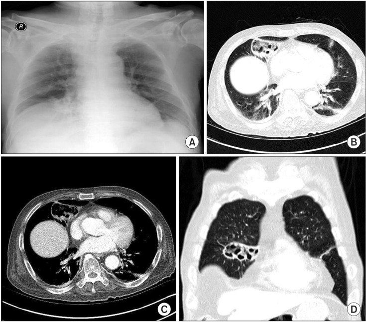
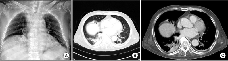
Pulmonary pneumatoceles are air-filled thin-walled spaces within the lung and are rare in adult cases of pneumonia. We report the case of a 74-year-old male who was admitted with a cough and sputum production. He had been treated with oral dexamethasone since a brain tumorectomy 6 months prior. Contrast-enhanced computed tomography (CT) of the chest revealed a large pneumatocele in the right middle lobe and peripheral pneumonic consolidation. Bronchoalveolar lavage was performed; cultures identified extended-spectrum β-lactamase (ESBL) producing Proteus mirabilis. A 4-week course of intravenous ertapenem was administered, and the pneumatocele with pneumonia resolved on follow-up chest CT. To the best of our knowledge, this is the first reported case of pulmonary pneumatocele caused by ESBL-producing P. mirabilis associated with pneumonia.
Keywords: Beta-Lactamase; Pneumonia; Proteus mirabilis.
Conflict of interest statement
Figures
Similar articles
-
Pneumatocele formation in a patient with Proteus mirabilis pneumonia.Postgrad Med J. 1985 Mar;61(713):255-7. doi: 10.1136/pgmj.61.713.255. Postgrad Med J. 1985. PMID: 3885205 Free PMC article.
-
Pneumatocele formation in adult pulmonary tuberculosis during antituberculous chemotherapy: a case report.Cases J. 2009 Sep 9;2:8570. doi: 10.4076/1757-1626-2-8570. Cases J. 2009. PMID: 19918388 Free PMC article.
-
Distribution and antimicrobial susceptibility profile of extended-spectrum β-lactamase-producing Proteus mirabilis strains recently isolated in Japan.Int J Antimicrob Agents. 2015 Feb;45(2):113-8. doi: 10.1016/j.ijantimicag.2014.06.005. Epub 2014 Aug 11. Int J Antimicrob Agents. 2015. PMID: 25182712
-
Carbapenems and piperacillin/tazobactam for the treatment of bacteremia caused by extended-spectrum β-lactamase-producing Proteus mirabilis.Diagn Microbiol Infect Dis. 2014 Nov;80(3):222-6. doi: 10.1016/j.diagmicrobio.2014.07.006. Epub 2014 Jul 26. Diagn Microbiol Infect Dis. 2014. PMID: 25139843
-
Antimicrobial susceptibility of clinical Enterobacteriaceae isolates at the emergency department in a regional hospital: A threat of extended spectrum beta-lactamase-producers among nursing home residents.J Microbiol Immunol Infect. 2016 Aug;49(4):584-90. doi: 10.1016/j.jmii.2015.10.001. Epub 2015 Nov 19. J Microbiol Immunol Infect. 2016. PMID: 26692184
Cited by
-
Pneumatocele formation in a fatal adult pneumonia patient coinfected with Streptococcus pyogenes emm-type 3 and influenza A: a case report.BMC Infect Dis. 2020 Nov 26;20(1):892. doi: 10.1186/s12879-020-05595-2. BMC Infect Dis. 2020. PMID: 33243155 Free PMC article.
-
Pneumatocele formation following COVID-19 pneumonia. Is there a role for surgical intervention?Asian Cardiovasc Thorac Ann. 2021 Dec 7;30(4):2184923211059866. doi: 10.1177/02184923211059866. Online ahead of print. Asian Cardiovasc Thorac Ann. 2021. PMID: 34874785 Free PMC article.
-
Proteus mirabilis: A rare cause of pneumonia, radiologically mimicking malignancy of the lung.Clin Case Rep. 2023 Sep 19;11(9):e7937. doi: 10.1002/ccr3.7937. eCollection 2023 Sep. Clin Case Rep. 2023. PMID: 37736480 Free PMC article.
-
Necrotizing tracheobronchitis associated with rheumatoid arthritis.Respir Med Case Rep. 2016 Nov 9;20:31-33. doi: 10.1016/j.rmcr.2016.11.006. eCollection 2017. Respir Med Case Rep. 2016. PMID: 27896063 Free PMC article.
References
-
- McGarry T, Giosa R, Rohman M, Huang CT. Pneumatocele formation in adult pneumonia. Chest. 1987;92:717–720. - PubMed
-
- Meyers HI, Jacobson G. Staphylococcal pneumonia in children and adults. Radiology. 1959;72:665–671. - PubMed
-
- Ceruti E, Contreras J, Neira M. Staphylococcal pneumonia in childhood: long-term follow-up including pulmonary function studies. Am J Dis Child. 1971;122:386–392. - PubMed
-
- Thapa BR, Kumar L, Mitra SK. Proteus mirabilis pneumonia with giant pneumatocele. Indian J Pediatr. 1987;54:593–597. - PubMed
LinkOut - more resources
Full Text Sources
Other Literature Sources