Single Nodular Pulmonary Amyloidosis: Case Report
- PMID: 26508930
- PMCID: PMC4620336
- DOI: 10.4046/trd.2015.78.4.385
Single Nodular Pulmonary Amyloidosis: Case Report
Abstract
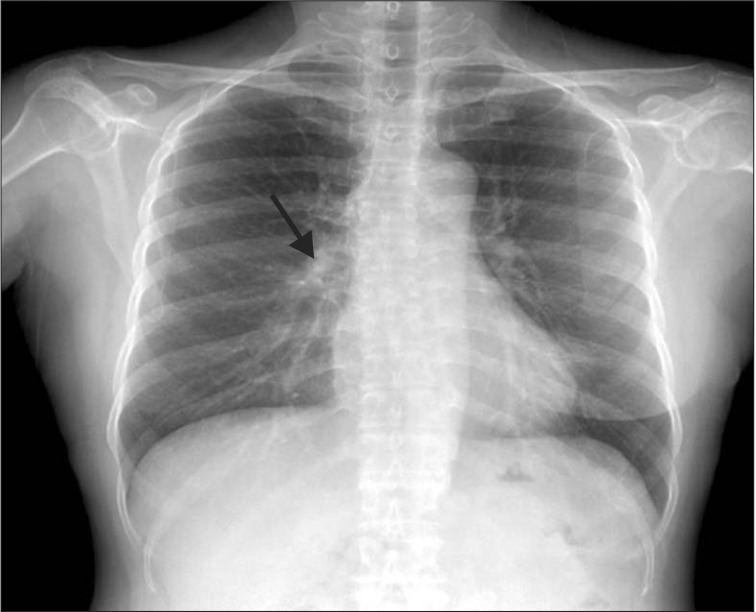
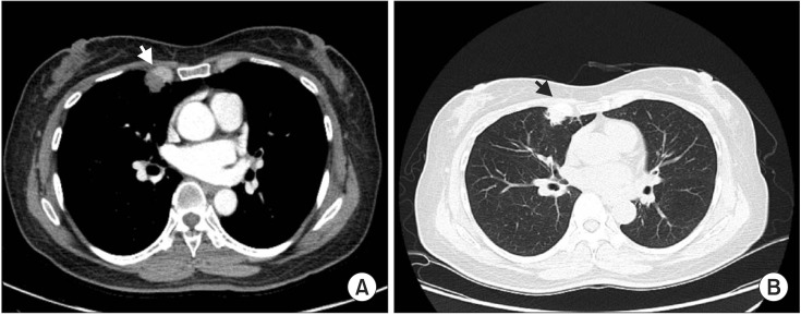
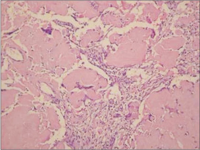
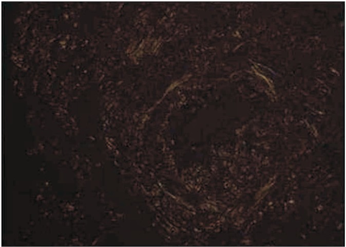
Amyloidosis is defined as the presence of extra-cellular deposits of an insoluble fibrillar protein, amyloid. The pulmonary involvement of amyloidosis is usually classified as tracheobronchial, parenchymal nodular, or diffuse alveolar septal. A single nodular lesion can mimic various conditions, including malignancy, pulmonary tuberculosis, and fungal infection. To date, only one case of nodular pulmonary amyloidosis has been reported in Korea, a case involving multiple nodular lesions. Here, we report and discuss the case of a patient having single nodular amyloidosis.
Keywords: Amyloidosis.
Conflict of interest statement
Figures
References
-
- Sipe JD, Benson MD, Buxbaum JN, Ikeda S, Merlini G, Saraiva MJ, et al. Amyloid fibril protein nomenclature: 2012 recommendations from the Nomenclature Committee of the International Society of Amyloidosis. Amyloid. 2012;19:167–170. - PubMed
-
- Utz JP, Swensen SJ, Gertz MA. Pulmonary amyloidosis. The Mayo Clinic experience from 1980 to 1993. Ann Intern Med. 1996;124:407–413. - PubMed
-
- Shin B, Ko J, Lee SS, Lim KS, Han JH, Chung MP, et al. A case of pulmonary amyloidosis mimicking lymphangitic lung carcinomatosis. Korean J Med. 2014;86:339–342.
-
- Glenner GG. Amyloid deposits and amyloidosis: the beta-fibrilloses (first of two parts) N Engl J Med. 1980;302:1283–1292. - PubMed
-
- Rajkumar SV, Gertz MA, Kyle RA. Primary systemic amyloidosis with delayed progression to multiple myeloma. Cancer. 1998;82:1501–1505. - PubMed
LinkOut - more resources
Full Text Sources
Other Literature Sources