. 2015:2015:424751.
doi: 10.1155/2015/424751. Epub 2015 Oct 5.

Oxidative Stress in Dilated Cardiomyopathy Caused by MYBPC3 Mutation

Affiliations

Oxidative Stress in Dilated Cardiomyopathy Caused by MYBPC3 Mutation

Thomas L Lynch 4th et al. Oxid Med Cell Longev. 2015.

Abstract

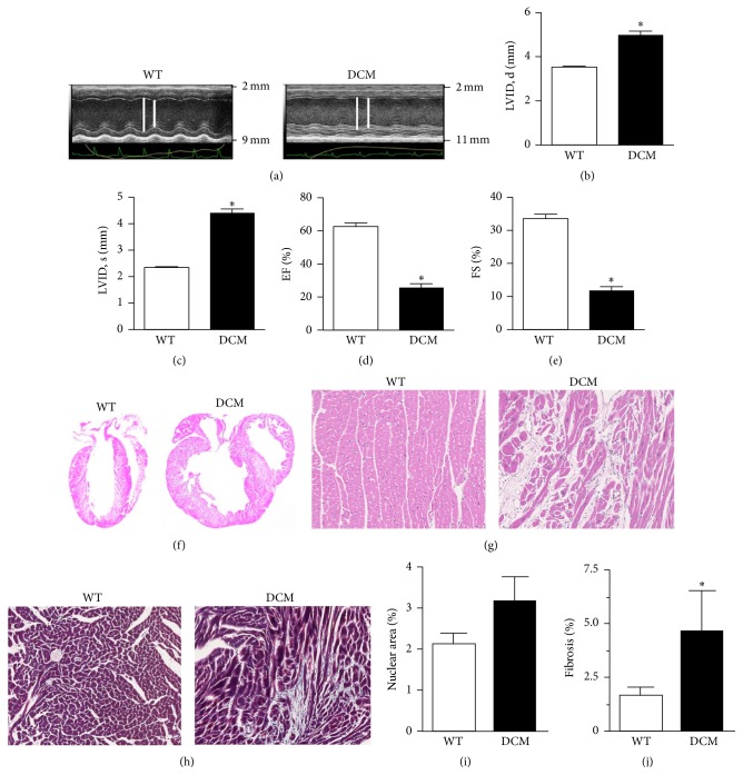
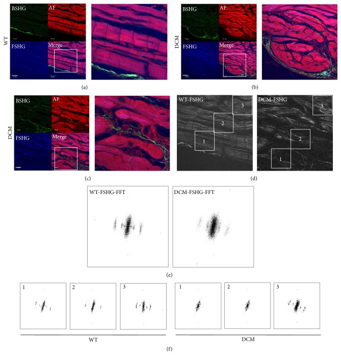
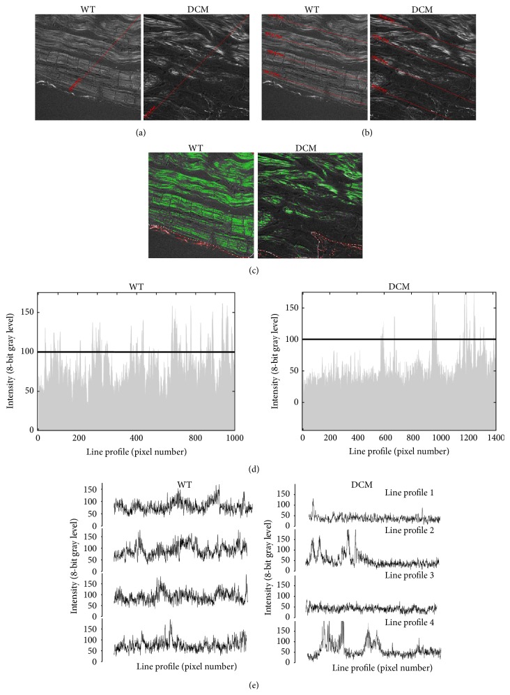
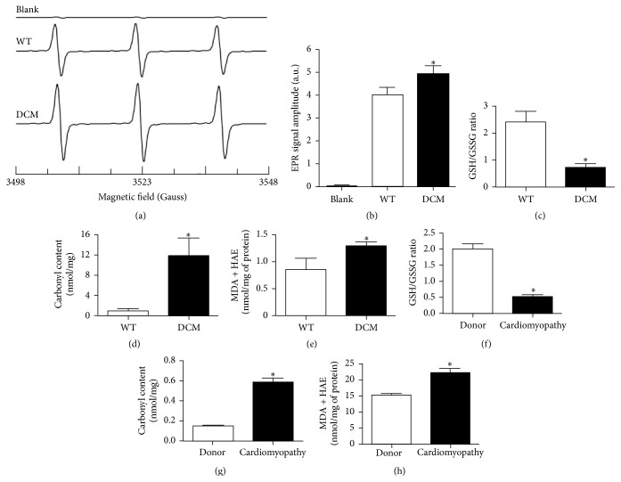
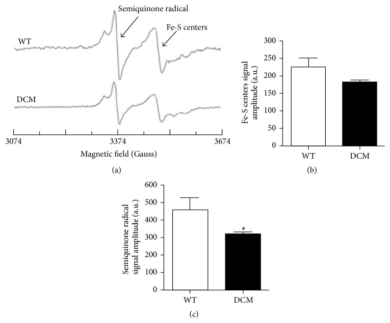
Cardiomyopathies can result from mutations in genes encoding sarcomere proteins including MYBPC3, which encodes cardiac myosin binding protein-C (cMyBP-C). However, whether oxidative stress is augmented due to contractile dysfunction and cardiomyocyte damage in MYBPC3-mutated cardiomyopathies has not been elucidated. To determine whether oxidative stress markers were elevated in MYBPC3-mutated cardiomyopathies, a previously characterized 3-month-old mouse model of dilated cardiomyopathy (DCM) expressing a homozygous MYBPC3 mutation (cMyBP-C((t/t))) was used, compared to wild-type (WT) mice. Echocardiography confirmed decreased percentage of fractional shortening in DCM versus WT hearts. Histopathological analysis indicated a significant increase in myocardial disarray and fibrosis while the second harmonic generation imaging revealed disorganized sarcomeric structure and myocyte damage in DCM hearts when compared to WT hearts. Intriguingly, DCM mouse heart homogenates had decreased glutathione (GSH/GSSG) ratio and increased protein carbonyl and lipid malondialdehyde content compared to WT heart homogenates, consistent with elevated oxidative stress. Importantly, a similar result was observed in human cardiomyopathy heart homogenate samples. These results were further supported by reduced signals for mitochondrial semiquinone radicals and Fe-S clusters in DCM mouse hearts measured using electron paramagnetic resonance spectroscopy. In conclusion, we demonstrate elevated oxidative stress in MYPBC3-mutated DCM mice, which may exacerbate the development of heart failure.


Figures

Figure 1
Figure 1
DCM hearts display contractile dysfunction, fibrosis, and myocardial disarray. (a) Representative parasternal long-axis M-mode echocardiographic images of WT and DCM hearts. Left ventricular internal diameter (LVID) is depicted by white lines (note: difference in scale between images). LVID at (b) peak diastole and (c) peak systole in WT and DCM hearts (n = 9 hearts, P < 0.001). Functional measurements derived from LVID measurements showing (d) ejection fraction (EF) and (e) fractional shortening (FS) in WT and DCM hearts (n = 9 hearts, P < 0.001). (f) H&E labeled whole hearts from WT and DCM animals. (g) H&E or (h) Masson's trichrome-labeled regions of interest (ROI) from WT and DCM hearts. (i) Quantification of cellularity based on nuclear area between WT (n = 5 hearts, 5 sections per heart) and DCM hearts (n = 4 hearts, 5 sections per heart). (j) Quantification of fibrosis (trichrome staining) area between WT (n = 4 hearts, 20 sections per heart) and DCM hearts (n = 4 hearts, 20 sections per heart; P < 0.0225). Data are represented as mean ± SEM. Significant differences (P < 0.05) between mean values were determined using Student's t-test.
Figure 2
Figure 2
Second harmonic generation (SHG) imaging demonstrates disorganized sarcomere pattern in DCM hearts. (a) The sarcomere pattern in WT hearts closer to the inner chamber is discernible, is well-organized, and has low levels of collagen (green) (scale bar, 133 μm). (b) In contrast, DCM hearts lack an organized sarcomere pattern (scale bar, 50 μm). (c) High magnification (HM) image of an area in (b), showing clear loss of sarcomeric structure in these swollen areas. Green channel is backward SHG (BSHG) depicting predominantly collagen fibers, red channel is autofluorescence (AF), and blue channel is forward directed SHG (FSHG) showing sarcomere pattern and their merge. (d) Fast Fourier Transformation (FFT) analysis of the sarcomere pattern in WT and DCM hearts from the inner chamber area to distal parts of the hearts. (e) FFT of the whole frames in (d), note the fuzziness in the high frequency pattern. However, the localized changes in sarcomere pattern could not be discerned while doing whole frame FFT analysis. (f) FFTs of regions of interest (1, 2, and 3) from the inner chamber of heart to distal part as depicted in highlighted areas in (d) showing the region specific disorganization in sarcomeric pattern. Note there is no difference in FFT pattern in location 3 of both WT and DCM hearts, although, in regions closer to the inner chamber (2) and especially in swollen regions (1), there is a substantial difference in DCM hearts but there are no discernible differences in WT. Images ((a)–(d)) are representative of multiple locations of 2 hearts (n = 2).
Figure 3
Figure 3
Loss of high frequency information (bands) directly indicates loss of typical sarcomere ladder structure in DCM hearts. Sarcomere fine structure in images from Figure 2(d) for DCM and WT heart sections is traced by drawing line intensity profiles across the cellular structures. (a) Loss of cellular structure in DCM (right) compared to WT (left) heart sections is indicated by drawing a single line from the inner chamber, which is (d) represented by line intensity profiles for both WT (left) and DCM (right) sections. (b) Loss of cellular structure is further indicated by multiple line profiles drawn left to right in DCM (right) compared to WT (left) heart sections, which is (e) represented by line intensity profiles (1–4) for both WT (left) and DCM (right) sections. (c) Sarcomere area imaging in WT (left) and DCM (right) hearts.
Figure 4
Figure 4
Increased oxidative stress in the hearts of DCM animals and cardiomyopathy patients. (a) Representative EPR spectra of CM measured at room temperature. Heart tissues (50 mg) were treated with CMH (1 mM) in Krebs buffer containing deferoxamine mesylate (25 μM) and incubated at 37°C in a water bath for 30 minutes. The reactive oxygen species oxidize CMH into CM. Blank samples are in the absence of heart tissue. (b) Summary data of CM (n = 3 hearts, P < 0.034 in DCM versus WT by Student's t-test, and blank shown for representation). No significant accumulation of CM was observed in the absence of heart tissues. (c) Comparison of GSH/GSSG ratios (n = 6 hearts), (d) carbonyl content (n = 6 hearts), and (e) MDA and HAE content (n = 8 hearts) in WT and DCM mouse heart tissue homogenates ( P < 0.05). (f) Comparison of GSH/GSSG ratios, (g) carbonyl content, and (h) MDA and HAE content in donor (n = 13 hearts) and cardiomyopathy (n = 14 hearts) human heart tissue homogenates ( P < 0.001). Data are represented as mean ± SEM. Significant differences (P < 0.05) between mean values were determined using Student's t-test.
Figure 5
Figure 5
Mitochondrial oxidative stress is augmented in the hearts of DCM animals. (a) Representative EPR spectra of heart tissue homogenates measured at 77 K. Semiquinone free radicals and Fe(III) from Fe-S centers signals are seen at g = 2.01 and g = 1.94, respectively. EPR signal amplitude was normalized against the weight of the heart and expressed as arbitrary units per gram. (b) Normalized EPR signal amplitude of the Fe-S centers (n = 3 hearts). (c) Normalized EPR signal amplitude of the semiquinone radical (n = 3 hearts, P < 0.026). Data are represented as mean ± SEM. Significant differences (P < 0.05) between mean values were determined using Student's t-test.

References

    1. Go A. S., Mozaffarian D., Roger V. L., et al Heart disease and stroke statistics—2014 update: a report from the American Heart Association. Circulation. 2014;129(3):e28–e292. doi: 10.1161/01.cir.0000441139.02102.80. - DOI - PMC - PubMed
    1. Morimoto S. Sarcomeric proteins and inherited cardiomyopathies. Cardiovascular Research. 2008;77(4):659–666. doi: 10.1093/cvr/cvm084. - DOI - PubMed
    1. Maron B. J., Maron M. S. Hypertrophic cardiomyopathy. The Lancet. 2013;381(9862):242–255. doi: 10.1016/S0140-6736(12)60397-3. - DOI - PubMed
    1. van Dijk S. J., Dooijes D., dos Remedios C., et al. Cardiac myosin-binding protein C mutations and hypertrophic cardiomyopathy: haploinsufficiency, deranged phosphorylation, and cardiomyocyte dysfunction. Circulation. 2009;119(11):1473–1483. doi: 10.1161/circulationaha.108.838672. - DOI - PubMed
    1. Marston S., Copeland O., Gehmlich K., Schlossarek S., Carrrier L. How do MYBPC3 mutations cause hypertrophic cardiomyopathy? Journal of Muscle Research and Cell Motility. 2012;33(1):75–80. doi: 10.1007/s10974-011-9268-3. - DOI - PubMed

Publication types

MeSH terms

LinkOut - more resources