. 2015 Sep 3:9:275-82.
doi: 10.1016/j.nicl.2015.08.014. eCollection 2015.

EEG resting state analysis of cortical sources in patients with benign epilepsy with centrotemporal spikes

Affiliations

EEG resting state analysis of cortical sources in patients with benign epilepsy with centrotemporal spikes

Azeez Adebimpe et al. Neuroimage Clin. .

Abstract

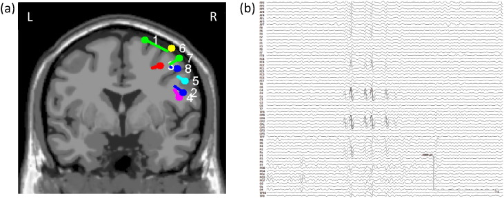
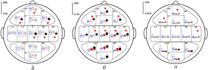
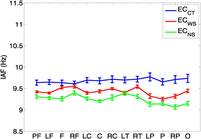
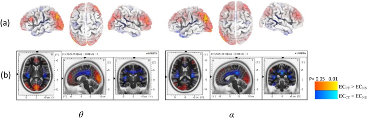
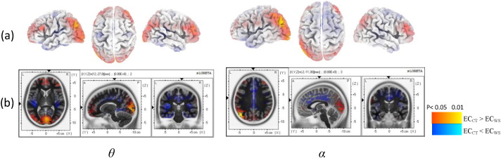
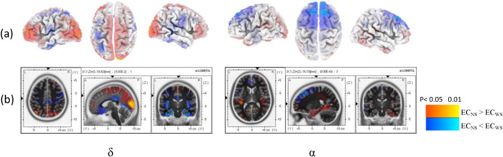
Benign epilepsy with centrotemporal spikes (BECTS) is the most common idiopathic childhood epilepsy, which is often associated with developmental disorders in children. In the present study, we analyzed resting state EEG spectral changes in the sensor and source spaces in eight BECTS patients compared with nine age-matched controls. Using high-resolution scalp EEG data, we assessed statistical differences in spatial distributions of EEG power spectra and cortical sources of resting state EEG rhythms in five frequency bands: δ (0.5-3.5 Hz), θ (4-8 Hz), α (8.5-13 Hz), β1 (13.5-20 Hz) and β2 (20.5-30 Hz) under the eyes-closed resting state condition. To further investigate the impact of centrotemporal spikes on EEG spectra, we split the EEG data of the patient group into EEG portions with and without spikes. Source localization demonstrated the homogeneity of our population of BECTS patients with a common epileptic zone over the right centrotemporal region. Significant differences in terms of both spectral power and cortical source densities were observed between controls and patients. Patients were characterized by significantly increased relative power in θ, α, β1 and β2 bands in the right centrotemporal areas over the spike zone and in the right temporo-parieto-occipital junction. Furthermore, the relative power in all bands significantly decreased in the bilateral frontal and parieto-occipital areas of patients regardless of the presence or absence of spikes in EEG segments. However, the spectral differences between patients and controls were more pronounced in the presence of spikes. This observation emphasized the impact of benign epilepsy on cortical source power, especially in the right centrotemporal regions. Spectral changes in bilateral frontal and parieto-occipital areas may also suggest alterations in the default mode network in BECTS patients.

Keywords: Benign epilepsy; Centrotemporal spikes; EEG; Source analysis; Spectral power.


Figures

Fig. 1
Fig. 1
(a) Dipole locations of the averaged spikes for patients, and (b) a sample EEG recording from patient 1.
Fig. 2
Fig. 2
Thirteen brain regions defined for statistical comparisons.
Fig. 3
Fig. 3
Average normalized spectral power maps (±standard deviation) in the sensor space under the eyes-closed condition for the control group (ECCT, left boxplots), patient group (ECNS, no spike condition, middle boxplots) and patient group (ECWS, with spike condition, right boxplots) in δ, θ and α bands. Significant differences between ECCT and ECNS/ECWS are shown in solid boxplots. Asterisks indicate statistically significant differences (p < 0.01) between ECNS and ECWS.
Fig. 4
Fig. 4
Individual alpha frequency (IAF) for all the 13 regions (see Fig. 2 for abbreviations).
Fig. 5
Fig. 5
Statistical maps of differences between cortical sources computed under the eyes-closed condition for the control group (ECCT, left boxplots) and the patient group (ECNS, no spike condition, right boxplots) in θ and α bands. The results have been projected onto the cortical layer of the realistic head model (a) and the MNI152 MRI (b). Color bars indicate significant differences between ECCT and ECNS, red (ECCT > ECNS) and blue (ECCT < ECNS).
Fig. 6
Fig. 6
Statistical maps of differences between cortical sources computed under the eyes-closed condition for the control group (ECCT, left boxplots) and patient group (ECWS, with spike condition, right boxplots) in θ and α bands. The results have been projected onto the cortical layer of the realistic head model (a) and the MNI152 MRI (b). Color bars indicate significant differences between ECCT and ECWS, red (ECCT > ECWS) and blue (ECCT < ECWS).
Fig. 7
Fig. 7
Statistical maps of differences between cortical sources computed under the no-spike (ECNS, left boxplots) and with-spike (ECWS, right boxplots) eyes-closed conditions in δ and α bands for the epileptic group. The results have been projected onto the cortical layer of the realistic head model (a) and the MNI152 MRI (b). Color bars indicate significant differences between ECNS and ECWS, red (ECns > ECWS) and blue (ECNS > ECWS).

Similar articles

Cited by

References

    1. Angelakis E., Lubar J.F., Stathopoulou S., Kounios J. Peak alpha frequency: an electroencephalographic measure of cognitive preparedness. Clin. Neurophysiol. Off. J. Int. Fed. Clin. Neurophysiol. 2004;115(4):887–897. 15003770 - PubMed
    1. Ansari S.F., Maher C.O., Tubbs R.S., Terry C.L., Cohen-Gadol A.A. Surgery for extratemporal nonlesional epilepsy in children: a meta-analysis. Childs Nerv. Syst. ChNS Off. J. Int. Soc. Pediatr. Neurosurg. 2010;26(7):945–951. 20013124 - PubMed
    1. Archer J.S., Abbott D.F., Waites A.B., Jackson G.D. fMRI “deactivation” of the posterior cingulate during generalized spike and wave. NeuroImage. 2003;20(4):1915–1922. 14683697 - PubMed
    1. Ay Y., Gokben S., Serdaroglu G., Polat M., Tosun A., Tekgul H., Solak U., Kesikci H. Neuropsychologic impairment in children with rolandic epilepsy. Pediatr. Neurol. 2009;41(5):359–363. 19818938 - PubMed
    1. Babiloni C., Pistoia F., Sarà M., Vecchio F., Buffo P., Conson M., Onorati P., Albertini G., Rossini P.M. Resting state eyes-closed cortical rhythms in patients with locked-in-syndrome: an EEG Study. Clin. Neurophysiol. Off. J. Int. Fed. Clin. Neurophysiol. 2010;121(11):1816–1824. 20541461 - PubMed

LinkOut - more resources