. 2015 Oct 28;10(10):e0140511.
doi: 10.1371/journal.pone.0140511. eCollection 2015.

Delineating the Tes Interaction Site in Zyxin and Studying Cellular Effects of Its Disruption

Affiliations

Delineating the Tes Interaction Site in Zyxin and Studying Cellular Effects of Its Disruption

Ermin Hadzic et al. PLoS One. .

Abstract

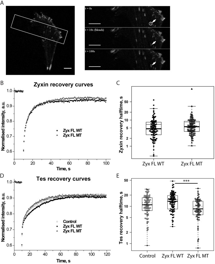
Focal adhesions are integrin-based structures that link the actin cytoskeleton and the extracellular matrix. They play an important role in various cellular functions such as cell signaling, cell motility and cell shape. To ensure and fine tune these different cellular functions, adhesions are regulated by a large number of proteins. The LIM domain protein zyxin localizes to focal adhesions where it participates in the regulation of the actin cytoskeleton. Because of its interactions with a variety of binding partners, zyxin has been proposed to act as a molecular scaffold. Here, we studied the interaction of zyxin with such a partner: Tes. Similar to zyxin, Tes harbors three highly conserved LIM domains of which the LIM1 domain directly interacts with zyxin. Using different zyxin variants in pull-down assays and ectopic recruitment experiments, we identified the Tes binding site in zyxin and showed that four highly conserved amino acids are crucial for its interaction with Tes. Based upon these findings, we used a zyxin mutant defective in Tes-binding to assess the functional consequences of abrogating the zyxin-Tes interaction in focal adhesions. Performing fluorescence recovery after photobleaching, we showed that zyxin recruits Tes to focal adhesions and modulates its turnover in these structures. However, we also provide evidence for zyxin-independent localization of Tes to focal adhesions. Zyxin increases focal adhesion numbers and reduces focal adhesion lifetimes, but does so independent of Tes. Quantitative analysis showed that the loss of interaction between zyxin and Tes affects the process of cell spreading. We conclude that zyxin influences focal adhesion dynamics, that it recruits Tes and that this interaction is functional in regulating cell spreading.


Conflict of interest statement

Competing Interests: The authors have declared that no competing interests exist.

Figures

Fig 1
Fig 1. Ectopic recruitment to mitochondria of Tes and Tes LIM by zyxin-mito variants.
(A) Schematic representation of the LIM domain proteins zyxin and Tes. The α-actinin binding site (AcBS) and the proline-rich region binding Ena/VASP family members (Pro-rich region) of zyxin are shown as well as the LIM domains. For Tes, the PET domain in the N-terminal part and the three LIM domains in the C-terminal part are depicted. In (B) and (C), Vero cells were transiently transfected with Zyx-mito variants and Tes FL-GFP, GFP-Tes LIM or GFP (control). Zyx FL WT-mito (B) and Zyx NT-mito (C) were labelled with an anti-zyxin antibody and Zyx LIM-myc-mito was labelled with an anti-myc antibody. Scale bar: 50 μm; the insets show a higher magnification of the outlined regions.
Fig 2
Fig 2. Determination of the minimal sequence of zyxin that interacts with Tes.
(A) Schematic representation of zyxin (1–572), fused to the mitochondrial targeting sequence (mito). The green box corresponds to the α-actinin binding site (AcBS), the four red boxes correspond to the four FPPPP motifs binding VASP, the yellow box represents the amino acids VGEI. Zyxin-mito variants are represented by lines showing their respective position in the full-length protein (in the same order as in the inset table). Their ability to recruit GFP-Tes LIM and GFP-Tes LIM1 on the surface of mitochondria in Vero cells is indicated in the table (nt: not tested). (B) and (C) Ectopic recruitment of GFP-Tes LIM1 by the zyxin variants Zyx 140-380-myc-mito, Zyx 51-140-myc-mito, Zyx 64-77-myc-mito and Zyx 51-63-myc-mito to the surface of the mitochondria in Vero cells. The different zyxin variants were labelled with an anti-myc antibody. Scale bar: 50 μm; the insets show a higher magnification of the outlined regions.
Fig 3
Fig 3. The sequence 51–63 contains 4 conserved amino acids VGEI important for the interaction between Tes and zyxin.
(A) Sequence alignment of the first 136 amino acids of human zyxin with orthologues (reference sequences with the indicated numbers were retrieved from the NCBI protein database for the indicated species, the zebrafish sequence was retrieved from Ensembl). Protein sequences were aligned with ClustalW2. The α-actinin binding site (green), the VGEI sequence (yellow) and the FPPPP motifs (red) are shown. (*) represents identical residues, (:) represents conserved substitutions and (·) represents semi-conserved substitutions in the alignment. (B) and (C) Ectopic recruitment of GFP-Tes LIM1 by zyxin variants to the surface of mitochondria in Vero cells. (B) Zyx 51-63-myc-mito and Zyx 51–63 MT-myc-mito sequences are indicated on top, both variants were labelled with an anti-myc antibody (top panels), (C) Zyx FL WT-mito and Zyx FL MT-mito were labelled with an anti-zyxin antibody (top panels). Bottom panels show GFP-Tes LIM1 signals. Scale bar: 50 μm; the insets show a higher magnification of the outlined regions.
Fig 4
Fig 4. The VGEI sequence in full-length zyxin is necessary for the interaction with Tes LIM1.
(A) and (C) expression levels of GFP-fusion proteins in HeLa cells were verified by Western blot using an anti-GFP antibody (A), an anti-zyxin antibody (C) and an anti-β-actin antibody (control) (A, C). (B) Western blot analysis of GST pull-down experiments performed with GST-Tes LIM1 immobilized on glutathione sepharose resin, and extracts of HeLa cells transfected with Zyx 51-63-GFP, Zyx 51–63 MT-GFP or GFP (negative control). The presence of zyxin-GFP variants was analyzed in bound “B” or non-bound “NB” fractions using an anti-GFP antibody. (D) Zyx FL WT-GFP and Zyx FL MT-GFP extracts from transfected HeLa cells, were analyzed in bound (B) and non-bound (NB) fractions using an anti-zyxin antibody. Note the presence of endogenous zyxin (zyxin endo) in the bound fraction.
Fig 5
Fig 5. The recruitment of Tes LIM1 to FAs depends on the VGEI sequence of zyxin.
(A) Wild-type and zyxin-null fibroblasts were transfected with GFP-Tes LIM1 or GFP (control). FAs were stained with an anti-vinculin antibody. (B) Zyxin-null fibroblasts were cotransfected with GFP-Tes LIM1 and DsRed, Zyx FL WT-DsRed or Zyx FL MT-DsRed. Scale bar: 50 μm, the insets show a higher magnification of the outlined regions.
Fig 6
Fig 6. Tes localizes to FAs in the absence of zyxin.
(A) Endogenous Tes was stained with an anti-Tes antibody in wild-type and zyxin-null fibroblasts and the FA marker vinculin was revealed with an anti-vinculin antibody. (B) Wild-type and zyxin-null fibroblasts were transfected with Tes FL-GFP and stained with an anti-vinculin antibody. Scale bar: 50 um; the insets show a higher magnification of the outlined regions. (C) and (D) Zyxin-null fibroblasts were cotransfected with Tes FL-GFP and the constructs DsRed (control), Zyx FL WT-DsRed or Zyx FL MT-DsRed. 24 hours after transfection, cells were fixed and stained for vinculin, and analyzed by immunofluorescence confocal microscopy. Measured intensities at FAs were then averaged for each cell. Bar plots indicate the intercellular means of measured intensities of Tes FL-GFP (C) and the means of the integrated fluorescence intensities of Zyx FL WT-DsRed and Zyx FL MT-DsRed (D) For all quantifications at least 25 cells and >1500 FAs per condition were analyzed. Error bars represent S.E.M. *** P<0.0001, only significant differences are indicated.
Fig 7
Fig 7. Dynamics of zyxin and Tes at FAs.
(A) A typical FRAP experiment with Zyx FL WT-GFP in zyxin-null fibroblasts. Left panel: A cell expressing Zyx FL WT-GFP. The white rectangle indicates the area selected for FRAP acquisition. Right panels represent the time-course of a FRAP experiment: prebleach image (top), first image after bleach (middle), 100 s after start of acquisition (bottom). The white circle outlines the region selected for the bleach. Scale bars: 10 μm. (B) Normalized and averaged Zyx FL WT-GFP (Zyx FL WT) and Zyx FL MT-GFP (Zyx FL MT) recovery curves obtained from four independent experiments (in total about 100 acquisitions per condition). (C) t ½ values of recovery halftimes of individual zyxin-GFP FRAP recoveries that were averaged in B are represented as Box-and-Whisker plots overlaid with data points. Thin lines inside boxes represent mean t ½ values. (D) Normalized and averaged Tes FL-GFP recovery curves in the presence of DsRed (Control), Zyx FL WT-DsRed (Zyx FL WT) or Zyx FL MT-DsRed (Zyx FL MT) obtained from four independent experiments (in total about 80 acquisitions per condition). (E) t ½ values of recovery halftimes of individual Tes FL-GFP FRAP recoveries that were averaged in D are represented as Box-and-Whisker plots overlaid with data points. Thin lines inside boxes represent mean t ½ values. *** P<0.0001, only significant differences are indicated.
Fig 8
Fig 8. Zyxin affects FA density in cells, FA morphology and FA lifecycle in a Tes-independent manner.
(A) Average FA size in the presence of DsRed (Control), Zyx FL WT-DsRed (Zyx FL WT) or Zyx FL MT-DsRed (Zyx FL MT). (B) Average number of FAs per μm2 of cell area in the presence of DsRed (Control), Zyx FL WT-DsRed (Zyx FL WT) or Zyx FL MT-DsRed (Zyx FL MT). In (A) and (B) measurements are based on vinculin staining with an anti-vinculin antibody and were first averaged per cell, barplots represent means ± S.E.M. of these values. For quantifications in A and B at least 25 cells and >1500 FAs per condition were analyzed. (C) Time-course panels representing different stages of FA lifecycle followed using mEmerald paxillin: FA de novo birth (upper panels), FA disassembly (middle panels), and stable FA existing throughout the observation time period (lower panels). Despite of having long lifetimes, these FAs can shift position and substantially change their phenotype. (D) Histogram representing the distribution of FA lifetimes in the presence of DsRed (Control), Zyx FL WT-DsRed (Zyx FL WT) or Zyx FL MT-DsRed (Zyx FL MT). 1023, 1618 and 1006 tracks accordingly were considered (see Materials and methods for details). (E) Fraction number of stable FAs (25, 65 and 51 tracks for Control, Zyx FL WT and Zyx FL MT respectively) presented as fractions relative to the average number of FAs per time frame. For quantifications in (D) and (E) at least 10 cells and >1000 FAs per condition were analyzed. *P<0.05, **P<0.01, ***P<0.0001, only significant differences are indicated.
Fig 9
Fig 9. Tes interaction with zyxin affects cell spreading.
Zyxin-null fibroblasts were transfected with GFP (Control), Zyx FL WT-GFP or Zyx FL MT-GFP. Spreading assays were performed as described in Materials and methods. Cell surfaces of individual transfected cells were determined 15 min, 30 min, 60 min, and 240 min after plating on fibronectin (20 μg/ml). Bar plots correspond to the means ± S.E.M. of 3 independent experiments. ** P<0.01, *** P<0.0001, only significant differences are indicated.

References

    1. Geiger B, Yamada KM. Molecular architecture and function of matrix adhesions. Cold Spring Harbor perspectives in biology. 2011;3(5). Epub 2011/03/29. 10.1101/cshperspect.a005033 - DOI - PMC - PubMed
    1. Gregory Call S, Brereton D, Bullard JT, Chung JY, Meacham KL, Morrell DJ, et al. A zyxin-nectin interaction facilitates zyxin localization to cell-cell adhesions. Biochemical and biophysical research communications. 2011;415(3):485–9. Epub 2011/11/08. 10.1016/j.bbrc.2011.10.099 . - DOI - PubMed
    1. Beckerle MC. Zyxin: zinc fingers at sites of cell adhesion. BioEssays: news and reviews in molecular, cellular and developmental biology. 1997;19(11):949–57. Epub 1997/12/12. 10.1002/bies.950191104 . - DOI - PubMed
    1. Hirata H, Tatsumi H, Sokabe M. Zyxin emerges as a key player in the mechanotransduction at cell adhesive structures. Communicative & integrative biology. 2008;1(2):192–5. Epub 2009/06/11. - PMC - PubMed
    1. Wojtowicz A, Babu SS, Li L, Gretz N, Hecker M, Cattaruzza M. Zyxin mediation of stretch-induced gene expression in human endothelial cells. Circulation research. 2010;107(7):898–902. Epub 2010/08/14. 10.1161/CIRCRESAHA.110.227850 . - DOI - PubMed

Publication types

MeSH terms

LinkOut - more resources