

Cervical vertebrae maturation index estimates on cone beam CT: 3D reconstructions vs sagittal sections
- PMID: 26509559
- PMCID: PMC5083889
- DOI: 10.1259/dmfr.20150162
Cervical vertebrae maturation index estimates on cone beam CT: 3D reconstructions vs sagittal sections
Abstract
Objectives: The aim of this study was to evaluate the performance of CBCT three-dimensional (3D) reconstructions and sagittal sections for estimates of cervical vertebrae maturation index (CVMI).
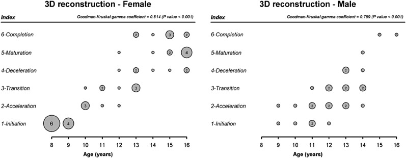
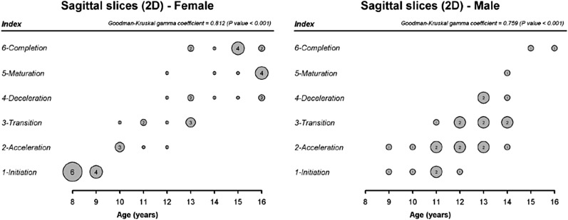
Methods: The sample consisted of 72 CBCT examinations from patients aged 8-16 years (45 females and 27 males) selected from the archives of two private clinics. Two calibrated observers (kappa scores: ≥0.901) interpreted the CBCT settings twice. Intra- and interobserver agreement for both imaging exhibition modes was analyzed by kappa statistics, which was also used to analyze the agreement between 3D reconstructions and sagittal sections. Correlations between cervical vertebrae maturation estimates and chronological age, as well as between the assessments by 3D reconstructions and sagittal sections, were analyzed using gamma Goodman-Kruskal coefficients (α = 0.05).
Results: The kappa scores evidenced almost perfect agreement between the first and second assessments of the cervical vertebrae by 3D reconstructions (0.933-0.983) and sagittal sections (0.983-1.000). Similarly, the agreement between 3D reconstructions and sagittal sections was almost perfect (kappa index: 0.983). In most divergent cases, the difference between 3D reconstructions and sagittal sections was one stage of CVMI. Strongly positive correlations (>0.8, p < 0.001) were found not only between chronological age and CVMI but also between the estimates by 3D reconstructions and sagittal sections (p < 0.001).
Conclusions: Although CBCT imaging must not be used exclusively for this purpose, it may be suitable for skeletal maturity assessments.
Keywords: Bone development; Orthodontics; biological indicators; cone beam computed tomography.
Figures


References
-
- O'Reilly MT, Yanniello GJ. Mandibular growth changes and maturation of cervical vertebrae—a longitudinal cephalometric study. Angle Orthod 1988; 58: 179–84. - PubMed
-
- Garcia-Fernandez P, Torre H, Flores L, Rea J. The cervical vertebrae as maturational indicators. J Clin Orthod 1998; 32: 221–5. - PubMed
-
- Baccetti T, Franchi L, McNamara JA, Jr. An improved version of cervical vertebral maturation (CVM) method for the assessment of mandibular growth. Angle Orthod 2002; 72: 316–23. - PubMed
MeSH terms
LinkOut - more resources
Full Text Sources
Other Literature Sources
Medical
