. 2015 Oct 16:6:8505.
doi: 10.1038/ncomms9505.

Structural basis for phosphatidylinositol-phosphate biosynthesis

Affiliations

Structural basis for phosphatidylinositol-phosphate biosynthesis

Oliver B Clarke et al. Nat Commun. .

Abstract

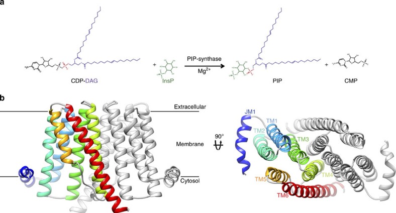
Phosphatidylinositol is critical for intracellular signalling and anchoring of carbohydrates and proteins to outer cellular membranes. The defining step in phosphatidylinositol biosynthesis is catalysed by CDP-alcohol phosphotransferases, transmembrane enzymes that use CDP-diacylglycerol as donor substrate for this reaction, and either inositol in eukaryotes or inositol phosphate in prokaryotes as the acceptor alcohol. Here we report the structures of a related enzyme, the phosphatidylinositol-phosphate synthase from Renibacterium salmoninarum, with and without bound CDP-diacylglycerol to 3.6 and 2.5 Å resolution, respectively. These structures reveal the location of the acceptor site, and the molecular determinants of substrate specificity and catalysis. Functional characterization of the 40%-identical ortholog from Mycobacterium tuberculosis, a potential target for the development of novel anti-tuberculosis drugs, supports the proposed mechanism of substrate binding and catalysis. This work therefore provides a structural and functional framework to understand the mechanism of phosphatidylinositol-phosphate biosynthesis.


Figures

Figure 1
Figure 1. Transmembrane architecture of a PIP synthase from R. salmoninarum.
(a) PIP synthases catalyse the transfer of a diacylglycerol-substituted phosphate group (purple/red) from the CDP-DAG donor to the inositol phosphate acceptor (green), generating PIP and CMP. (b) Structure of the RsPIPS-Δ6N homodimer in ribbon representation viewed from two orthogonal orientations (in the plane of the membrane on the left; towards the cytosol down the dimer axis on the right). One protomer is coloured grey, and the helices of the other are depicted in spectral colouring, from blue (JM1) to red (TM6). The Af2299 extramembrane domain used to facilitate crystallization is not shown here.
Figure 2
Figure 2. A large interfacial cavity contains the active site of RsPIPS.
(a) The structure of RsPIPS-Δ6N is shown in ribbon representation, with one protomer coloured grey and the other coloured by the Kyte–Doolitle hydrophobicity scale, from −4.5 (most polar, light blue) to 4.5 (most hydrophobic, orange). Two orthogonal representations are shown, on the left is a view in the plane of the membrane and on the right is a view from the cytosol along the dimer axis. A transparent purple surface (calculated using the 3V server35) delineates the borders of the interfacial cavity, which contains three subregions as follows: 1, the inositol phosphate acceptor-binding pocket; 2, the nucleotide-binding pocket between TM2 and TM3; and 3, a hydrophobic groove between TM2 and JM1. (b) Detail of the active site viewed in the plane of the membrane, with side chains that contact the bound Mg2+ and formula image 
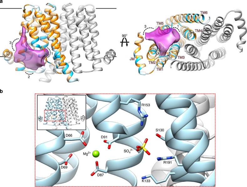
ions labelled and depicted in stick representation.
Figure 3
Figure 3. CDP-DAG binding to RsPIPS-FL.
The structure of RsPIPS-FL, depicted in ribbon representation and coloured as per Fig. 1, is shown in three different views (ac) superimposed on a transparent ‘Stromboli black' spacefill model (upper panels), with matching magnified insets of boxed regions below (lower panels). CDP-DAG is depicted in purple and Mg2+ ions in light green. Side chains that contact CDP-DAG or Mg2+ are shown in stick representation.
Figure 4
Figure 4. A homology model of MtPIPS, with functional characterization of selected point mutants.
(a) The MtPIPS homology model is shown in ribbon representation with one protomer in grey and the other in light blue, from two views, on the left viewed in the plane of the membrane and on the right from the cytosol, along the dimer axis. The substrates, CDP-DAG (purple) and inositol phosphate (black), are modelled based on the structures of RsPIPS-FL (in complex with CDP-DAG) and RsPIPS-Δ6N (with bound SO42−). The homology model was generated using the Phyre2 server, in one-to-one threading mode using the sequence of MtPIPS (Uniprot accession: P9WPG7) as the target and the structure of RsPIPS-FL (with the Af2299 extramembrane domain excised) as the template. Selected residues which are predicted to participate in either inositol phosphate binding (R155, R195, S132, K135), CDP-DAG binding (P153, M69, D31), neither (L70), or catalysis (D93) are shown with side chains in stick representation and coloured as in (b), where the activity of point mutants at these positions for the MtPIPS-FL construct is shown compared to wild-type MtPIPS-FL. (c) formula image 
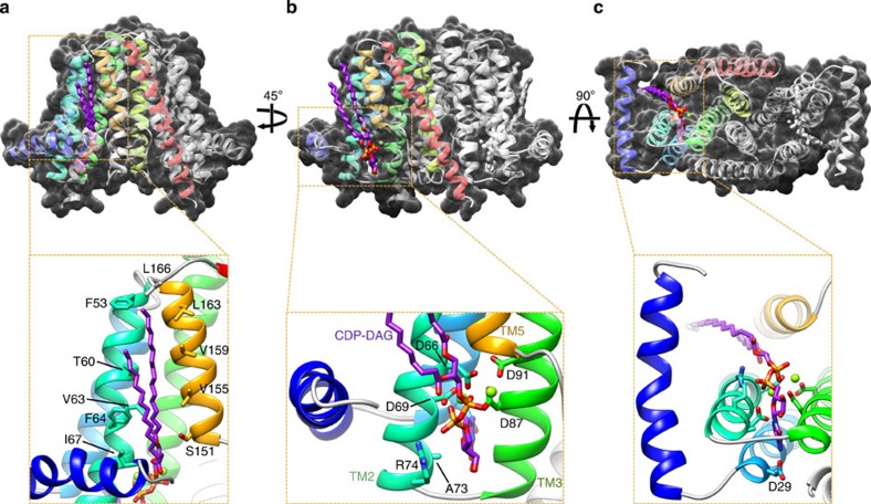
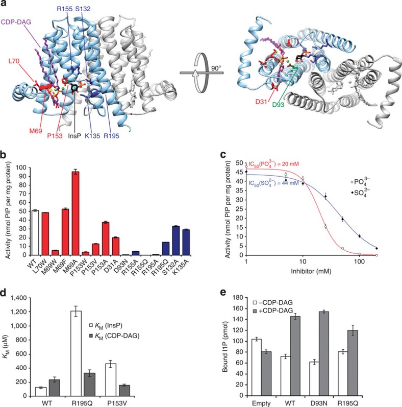
(closed diamonds) and formula image (open circles) inhibit the activity of MtPIPS-FL with half-inhibitory concentrations of 44 and 22 mM, respectively. (d) KM of MtPIPS-FL WT, R195Q and P153V for inositol phosphate (InsP; white) and CDP-DAG (grey). InsP, L-myo-inositol-1-phosphate (inositol phosphate). (e) Quantification of bound L-myo-[14C]inositol-1-phosphate after incubation of liposomes containing 9 μg MtPIPS-FL (WT, D93N, R195Q or empty liposome control) in the presence and absence of 200 μM CDP-DAG with 40 μM L-myo-[14C]inositol-1-phosphate. Measurement errors were quantified as s.e.m. (n=3).

References

    1. Umesiri F. E., Sanki A. K., Boucau J., Ronning D. R. & Sucheck S. J. Recent advances toward the inhibition of mAG and LAM synthesis in Mycobacterium tuberculosis. Med. Res. Rev. 30, 290–326 (2010). - PubMed
    1. Angala S. K., Belardinelli J. M., Huc-Claustre E., Wheat W. H. & Jackson M. The cell envelope glycoconjugates of Mycobacterium tuberculosis. Crit. Rev. Biochem. Mol. Biol. 49, 1–39 (2014). - PMC - PubMed
    1. Fischl A. S. & Carman G. M. Phosphatidylinositol biosynthesis in Saccharomyces cerevisiae: purification and properties of microsome-associated phosphatidylinositol synthase. J. Bacteriol. 154, 304–311 (1983). - PMC - PubMed
    1. Morii H., Ogawa M., Fukuda K., Taniguchi H. & Koga Y. A revised biosynthetic pathway for phosphatidylinositol in Mycobacteria. J. Biochem. 148, 593–602 (2010). - PubMed
    1. Morii H., Kiyonari S., Ishino Y. & Koga Y. A novel biosynthetic pathway of archaetidyl-myo-inositol via archaetidyl-myo-inositol phosphate from CDP-archaeol and D-glucose 6-phosphate in methanoarchaeon Methanothermobacter thermautotrophicus cells. J. Biol. Chem. 284, 30766–30774 (2009). - PMC - PubMed

Publication types

MeSH terms

Substances

LinkOut - more resources