. 2016 Jan;228(1):26-46.
doi: 10.1111/joa.12400. Epub 2015 Oct 29.

Innervation of the rabbit cardiac ventricles

Affiliations

Innervation of the rabbit cardiac ventricles

Neringa Pauziene et al. J Anat. 2016 Jan.

Abstract

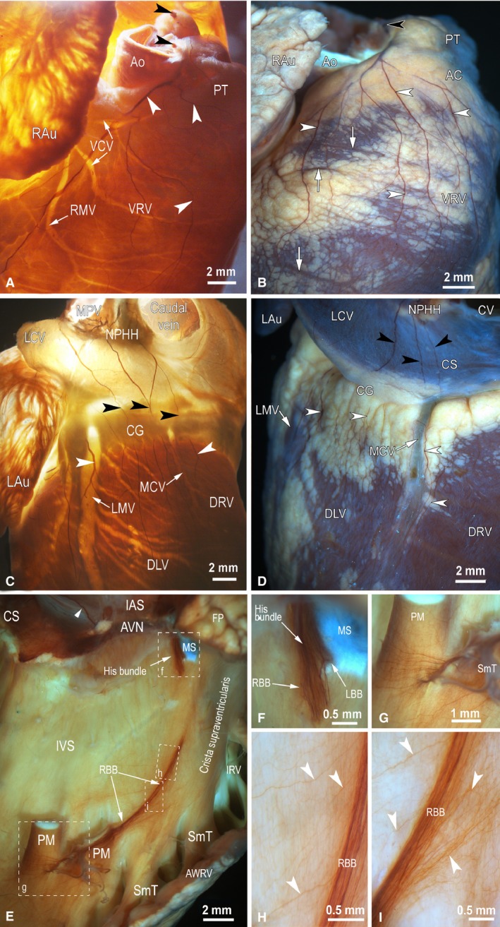
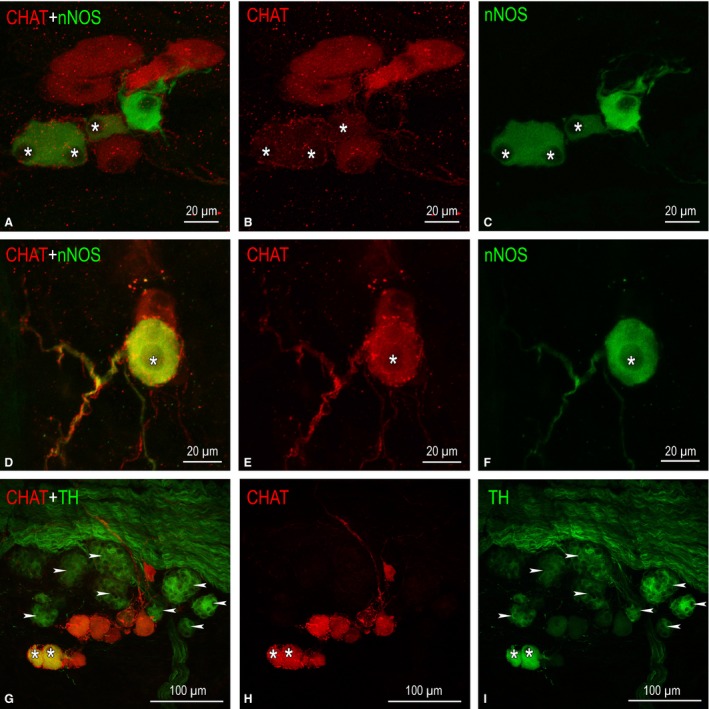
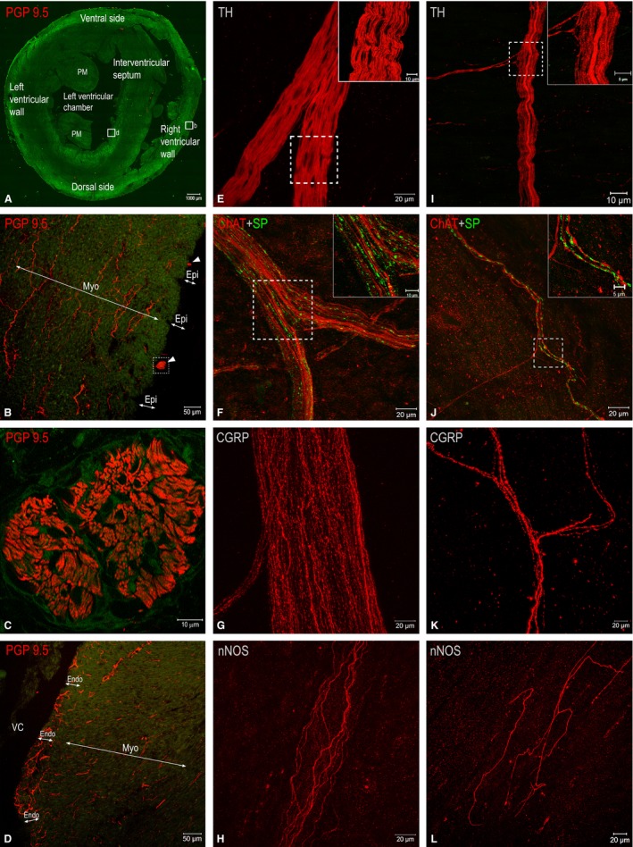
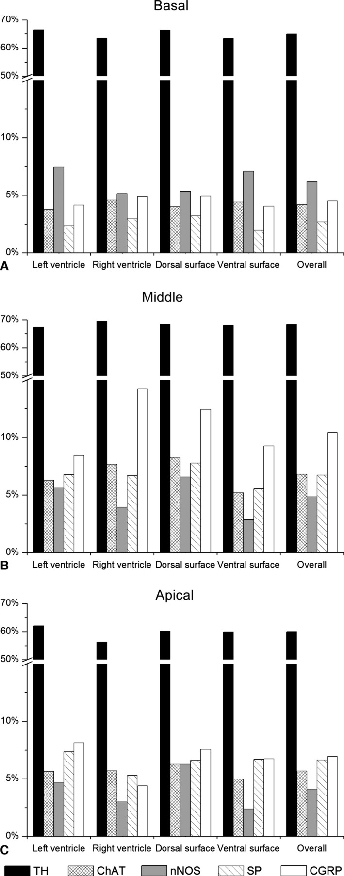
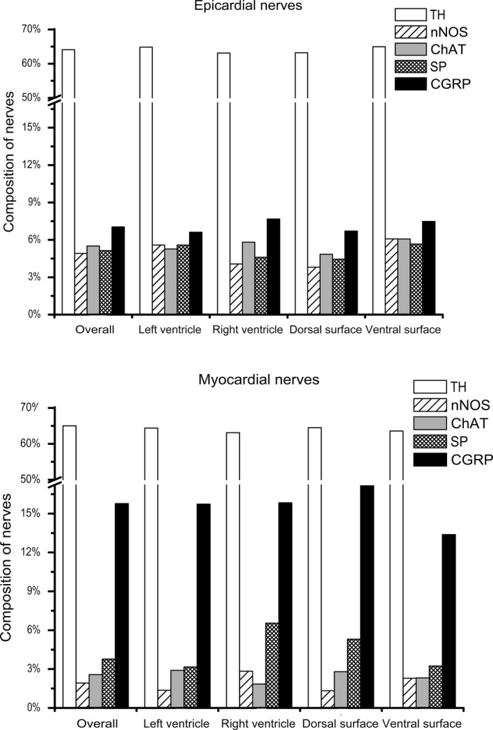
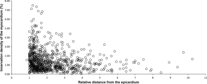
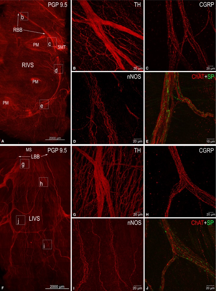
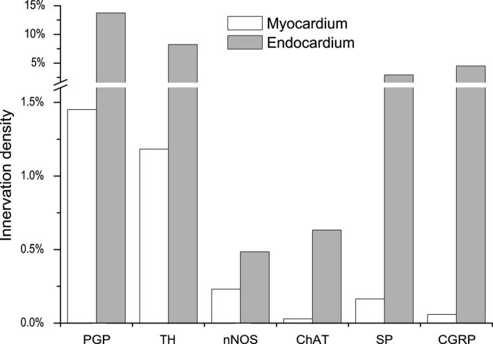
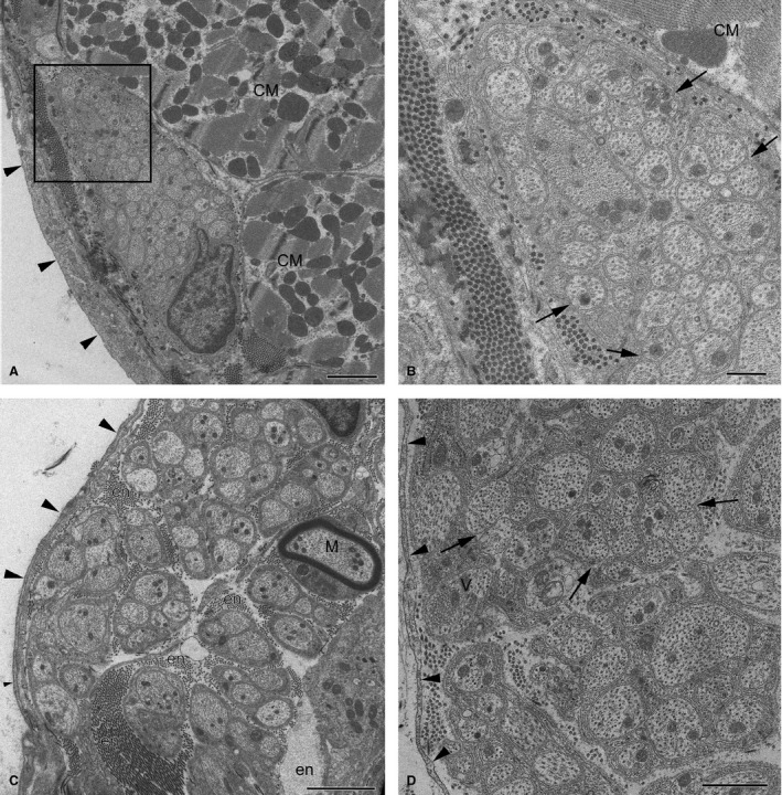
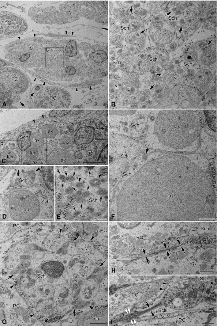
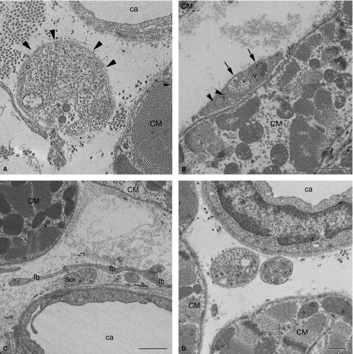
The rabbit is widely used in experimental cardiac physiology, but the neuroanatomy of the rabbit heart remains insufficiently examined. This study aimed to ascertain the architecture of the intrinsic nerve plexus in the walls and septum of rabbit cardiac ventricles. In 51 rabbit hearts, a combined approach involving: (i) histochemical acetylcholinesterase staining of intrinsic neural structures in total cardiac ventricles; (ii) immunofluorescent labelling of intrinsic nerves, nerve fibres (NFs) and neuronal somata (NS); and (iii) transmission electron microscopy of intrinsic ventricular nerves and NFs was used. Mediastinal nerves access the ventral and lateral surfaces of both ventricles at a restricted site between the root of the ascending aorta and the pulmonary trunk. The dorsal surface of both ventricles is supplied by several epicardial nerves extending from the left dorsal ganglionated nerve subplexus on the dorsal left atrium. Ventral accessing nerves are thicker and more numerous than dorsal nerves. Intrinsic ventricular NS are rare on the conus arteriosus and the root of the pulmonary trunk. The number of ventricular NS ranged from 11 to 220 per heart. Four chemical phenotypes of NS within ventricular ganglia were identified, i.e. ganglionic cells positive for choline acetyltransferase (ChAT), neuronal nitric oxide synthase (nNOS), and biphenotypic, i.e. positive for both ChAT/nNOS and for ChAT/tyrosine hydroxylase. Clusters of small intensely fluorescent cells are distributed within or close to ganglia on the root of the pulmonary trunk, but not on the conus arteriosus. The largest and most numerous intrinsic nerves proceed within the epicardium. Scarce nerves were found near myocardial blood vessels, but the myocardium contained only a scarce meshwork of NFs. In the endocardium, large numbers of thin nerves and NFs proceed along the bundle of His and both its branches up to the apex of the ventricles. The endocardial meshwork of fine NFs was approximately eight times denser than the myocardial meshwork. Adrenergic NFs predominate considerably in all layers of the ventricular walls and septum, whereas NFs of other neurochemical phenotypes were in the minority and their amount differed between the epicardium, myocardium and endocardium. The densities of NFs positive for nNOS and ChAT were similar in the epicardium and endocardium, but NFs positive for nNOS in the myocardium were eight times more abundant than NFs positive for ChAT. Potentially sensory NFs positive for both calcitonin gene-related peptide and substance P were sparse in the myocardial layer, but numerous in epicardial nerves and particularly abundant within the endocardium. Electron microscopic observations demonstrate that intrinsic ventricular nerves have a distinctive morphology, which may be attributed to remodelling of the peripheral nerves after their access into the ventricular wall. In conclusion, the rabbit ventricles display complex structural organization of intrinsic ventricular nerves, NFs and ganglionic cells. The results provide a basic anatomical background for further functional analysis of the intrinsic nervous system in the cardiac ventricles.

Keywords: CGRP; PGP 9.5; SP; choline acetyltransferase; electron microscopy; heart ventricles; innervation; nNOS; rabbit; tyrosine hydroxylase.


Figures

Figure 1
Figure 1
Macrophotographs to illustrate the distribution of epicardial nerves (white arrowheads) on rabbit ventricles (A–D) and endocardial nerves on the right side of the interventricular septum (E–I) stained histochemically for AChE. (A, B) The anterolateral view of the right ventricle; (C, D) the posterodorsal view of both ventricles; (E–I) the lateral view of the right side of the interatrial (IAS) and interventricular (IVS) septa. Cardiac ventricles in (A) and (C) are illuminated by transmission light, in (B) and (D) by reflecting light, and in (E)–(I) a combination of transmission and reflecting lights was applied to optimally illuminate the neural structures distributed in cardiac septa. Black arrowheads point to nerves that accessed the ventricles at the site between the roots of the ascending aorta and pulmonary trunk (A, B) and the nerves that extend towards the dorsal side of ventricles from the left atrium (C, D). White arrowheads in (A)–(D) indicate epicardial nerves that extend from the basal level of cardiac ventricles towards the apical level of ventricles and, in (E), an endocardial nerve of the interatrial septum that accesses the atrioventricular nodal region from the posterodorsal side of the interatrial septum. White arrows in (A)–(D) show the beds of some coronary vessels. The boxed areas in (E) are enlarged as (F)–(I). Abbreviations: AC, arterial conus; Ao, the root of ascending aorta; AVN, atrioventricular node; AWRV, anterior wall of the right ventricle; CG, coronary groove; CS, coronary sinus; CV, caudal vein; DLV, dorsal surface of left ventricle; DRV, dorsal surface of right ventricle; FP, fat pad at the root of ascending aorta; IAS, interatrial septum; IRV, infundibulum of right ventricle; IVS, interventricular septum; LAu, left auricle; LBB, left branch of the His bundle; LCV, left cranial vein; LMV, left marginal vein; MCV, middle cardiac vein; MPV, middle pulmonary vein; MS, membranous septum; NPHH, ganglionated nerve plexus in the limits of heart hilum at the root of middle pulmonary vein; PM, papillary muscle; PT, pulmonary trunk; RAu , right auricle; RBB, right branch of the His bundle; RMV, right marginal vein; SmT, septomarginal trabecula; VCV, ventral cardiac veins; VRV, ventral surface of right ventricle.
Figure 2
Figure 2
NS distributed on the conus arteriosus and the root of the pulmonary trunk of rabbit ventricles. (A–F) Small epicardial ganglia found on the arterial cone that contain NS positive for choline acetyltransferase (ChAT; in red), for neuronal nitric oxide synthase (nNOS; in green), and biphenotypic somata (labelled by asterisks), i.e. simultaneously positive for ChAT and nNOS. In (A) and (C), note the comparatively small size of NS positive for nNOS; (D–F) ganglion of two neurons, one of which is ChAT‐positive and another biphenotypic with positivity for nNOS (*). (G–I) A little ganglion located nearby a thick nerve that accessed cardiac ventricles throughout the arterial part of the heart hilum. NS positive for ChAT are shown in red, for tyrosine hydroxylase (TH) in green, and biphenotypic somata, i.e. simultaneously positive for ChAT and TH, are labelled with asterisks. Numerous SIF cells strongly positive for TH (labelled arrowheads) and clustered into tiny groups are located in between NS and a nerve containing plentiful NFs positive for TH.
Figure 3
Figure 3
(A–D) Fluorescent microphotographs to illustrate transverse sections through rabbit cardiac ventricles (A) and the morphological patterns of nerves (white arrowheads) and NFs in transversely sectioned epicardial (B), myocardial (B, D) and endocardial (D) layers. The boxed areas in (A) are enlarged as (B) and (D), respectively. The boxed area in (B) is enlarged as (C), which demonstrates one of the thick epicardial nerves (white arrowheads in B) that is full of NFs immunohistochemically positive for PGP 9.5. (E–L) Laser‐scanning (confocal) microphotographs from whole‐mount preparations of the walls of rabbit ventricles demonstrating both epicardial (E–H) and endocardial (I–L) nerves that involve NFs positive for tyrosine hydroxylase (TH), choline acetyltransferase (ChAT), substance P (SP), calcitonin gene‐related peptide (CGRP) and neuronal nitric oxide synthase (nNOS). The boxed areas in (E), (F), (I) and (J) are enlarged as insets on the right upper corner. Abbreviations: Endo, endocardium; Epi, epicardium; Myo, myocardium; PM, papillary muscle; VC, ventricular chamber.
Figure 4
Figure 4
Distribution of NFs positive for tyrosine hydroxylase (TH), choline acetyltransferase (ChAT), neuronal nitric oxide synthase (nNOS), substance P (SP) and calcitonin gene‐related peptide (CGRP) within transversely sectioned epicardial nerves sampled in different locations at the basal (A), middle (B) and apical (C) levels of rabbit ventricles.
Figure 5
Figure 5
Distribution of NFs positive for tyrosine hydroxylase (TH), choline acetyltransferase (ChAT), neuronal nitric oxide synthase (nNOS), substance P (SP) and calcitonin gene‐related peptide (CGRP) within transversely sectioned epicardial and myocardial nerves sampled in different locations of rabbit ventricles.
Figure 6
Figure 6
Scatter diagram of the NF density in transversely sectioned myocardium vs. their distance from the centre of the cardiac ventricle. NF density is the highest subepicardially, and decreases gradually towards the endocardium.
Figure 7
Figure 7
Laser‐scanning (confocal) microphotographs to illustrate morphological (A, F) and neurochemical (B–E, G–J) patterns of endocardial nerves distributed along the AVCA on the right (A) and left (F) sides of the interventricular septum. (A) and (F) display mosaic images that were automatically assembled with the aid of a laser‐scanning microscope from numerous microphotographs of tissue samples immunofluorescent for the antigen PGP 9.5. The boxed areas in (A) and (F) indicate the approximate positions on the right (RIVS) and the left (LIVS) sides of the interventricular septum, from which endocardial nerves with immunohistochemically different NFs are correspondingly shown in (B)–(E), and (G) and (H). Abbreviations in the upper right corner indicate the antigens for which endocardial NFs were positive following the relevant immunohistochemical reactions. Abbreviations: CGRP, calcitonin gene‐related peptide; ChAT, choline acetyltransferase; LBB, left branch of the His bundle; LIVS, left side of interventricular septum; MS, membranous septum; nNOS, neuronal nitric oxide synthase; PM, papillary muscles; RBB, right branch of the His bundle; RIVS, right side of interventricular septum; SMT, septomarginal trabecula; SP, substance P; TH, tyrosine hydroxylase.
Figure 8
Figure 8
Comparative density of NFs positive for tyrosine hydroxylase (TH), choline acteyltransferase (ChAT), neuronal nitric oxide synthase (nNOS), substance P (SP) and calcitonin gene‐related peptide (CGRP) within the transversely sectioned myocardium and endocardium of rabbit ventricles.
Figure 9
Figure 9
Electron micrographs of intrinsic nerves in rabbit ventricles. (A) A profile of a small epicardial nerve; (B) the enlarged image of the boxed area (A). Note the oblong profile of the nerve with densely packed axons, which is located beneath the mesothelial cell layer (arrows) and is sheathed by dense collagen fibres with fibroblast processes instead of perineurium. Arrowheads in (B) point to groups of axons cuddled together in a polyaxonal pocket of Schwann cell. (C) A thick fibroblast‐ensheathed myocardial nerve from the septomarginal trabecula. Note the singular myelinated NF (M). (D) A profile of an endocardial nerve ensheathed by fibroblasts (arrowheads) with unmyelinated NFs that cuddle axons together in a polyaxonal pocket (arrows). Other abbreviations: CM, profiles of cardiomyocytes; en, endoneurium. Scale bars: 2 μm (A and C); 1 μm (D); 500 nm (B).
Figure 10
Figure 10
Electron micrographs of endocardial nerves related topographically to the AVCA in the interventricular septum. (A) Low‐magnification image illustrating the profiles of two thin nerves (N) that were found regularly between Purkinje cells (PC). The lower nerve profile is partly ensheathed by fibroblast processes (arrowheads), but the upper one has no sheath at all. In the lower left corner of the image, note the profiles of two NFs with several tiny axons (arrows) that are situated near Purkinje cells. The boxed area (b) in (A) is enlarged as (B), which in detail demonstrates unusual NFs with heterogeneous axon populations. Some axons contain varicosities with neurotransmitter vesicles (v) that are absent in the majority of axons. Note the greatly varying diameter of axons and the abundant groups of axons (arrows) cuddled together in polyaxonal pockets of Schwann cell (Sch). (C) A part of a profile of the endocardial nerve that was located in the bundle of His at the most superior site of the interventricular septum. Note the triple‐layered fibroblast cell processes (arrowheads) that surround the NFs of the nerve. NFs in this nerve enclose enormously large axons (lax) together with normal (ax) and exceptionally thin axons, which are poorly seen due to insufficient magnification of the electron micrograph. The areas with tiny axons (arrows) are boxed and enlarged as (D)–(F). Note that some tiny axons (arrows) are cuddled together in a polyaxonal pocket of a Schwann cell (Sch). (D–F) Enlarged areas from (C) exhibiting diversity of axon diameters in the NF. (G) A profile of a typical large NF situated in between Purkinje cells (PC) of the right branch of the bundle of His at the middle part of the interventricular septum. Note the heterogeneous diameters of axons, i.e. some axons are comparatively large and contain vesicles (v), but others (arrows) are obviously smaller, filled with condensed axoplasm and cuddled in polyaxonal pockets of a Schwann cell (Sch). The axons with neurotransmitter vesicles (v) are not covered by Schwann cell processes and they are open (arrowheads) to the side of the Purkinje cells (PC). Abbreviation: ca, a part of the capillary lumen. (H) Electron micrograph of a longitudinally sectioned tiny axon (arrows) located between two Purkinje cells (PC) that expands to its varicosity (ax1) and contacts with the varicosity of another axon (ax2). The boxed area in (H) is enlarged as (I). (I) Enlarged image of the boxed area of (H), which in detail demonstrates two synaptic sites between both the axon (ax1) and the Purkinje cell (PC, arrowheads), and between two varicose axons (ax1 and ax2). Note the Schwann cell processes (Sch) that do not cover the axon (ax1) from the side of Purkinje cells (PC, two arrowheads). The axon diameter is only 0.1 μm (black arrows) in the intervals without neurotransmitter vesicles, and expands up to 0.2 μm at the interval filled with vesicles (white arrows). Scale bars: 5 μm (A and C); 2 μm (H); 1 μm (B, D, F and G); 500 nm (E and I).
Figure 11
Figure 11
Electron micrographs of myocardial NFs in the rabbit ventricles. (A) Unmyelinated NF with 12 axons (labelled with Arabic numerals) in contact with each other and enclosed by a Schwann cell from just one side (arrowheads) near a capillary (ca) and a cardiomyocyte (CM). Note that one of the axons contains neurotransmitter vesicles (V). (B) Unmyelinated NF with few axons near a cardiomyocyte (CM). Note that two axons with transmitter vesicles (V) are separated from a cardiomyocyte by a Schwann cell, but are open (arrows) to the more distant myocyte (CM) seen in the upper left corner of the panel. Some axons are significantly smaller in diameter than others, and are in contact with each other (arrowheads). (C) Two singular axons near a capillary (ca) are separated from a cardiomyocyte (CM) by fibroblast processes (fb). One axon (ax) is only in part closed by a Schwann cell (Sch), the other one (ax) is completely unenveloped. (D) Terminal axon (ax) filled with neurotransmitter vesicles without any Schwann cells close to cardiomyocytes (CM). Scale bar: 1 μm (A–C); 500 nm (D).

References

    1. Anderson RH (1972a) The disposition, morphology and innervation of cardiac specialized tissue in the guinea‐pig. J Anat 111, 453–468. - PMC - PubMed
    1. Anderson RH (1972b) Histologic and histochemical evidence concerning the presence of morphologically distinct cellular zones within the rabbit atrioventricular node. Anat Rec 173, 7–24. - PubMed
    1. Armour JA (2010) Functional anatomy of intrathoracic neurons innervating the atria and ventricles. Heart Rhythm 7, 994–996. - PubMed
    1. Armour JA, Murphy DA, Yuan BX, et al. (1997) Gross and microscopic anatomy of the human intrinsic cardiac nervous system. Anat Rec 247, 289–298. - PubMed
    1. Atkinson CJ, Santer RM (1999) Quantitative studies on myelinated and unmyelinated nerve fibres in the interatrial septal region of aged rat hearts. J Auton Nerv Syst 77, 172–176. - PubMed

Publication types

Substances

LinkOut - more resources