. 2015 Oct 29:5:15603.
doi: 10.1038/srep15603.

Generation of a miniature pig disease model for human Laron syndrome

Affiliations

Generation of a miniature pig disease model for human Laron syndrome

Dan Cui et al. Sci Rep. .

Abstract

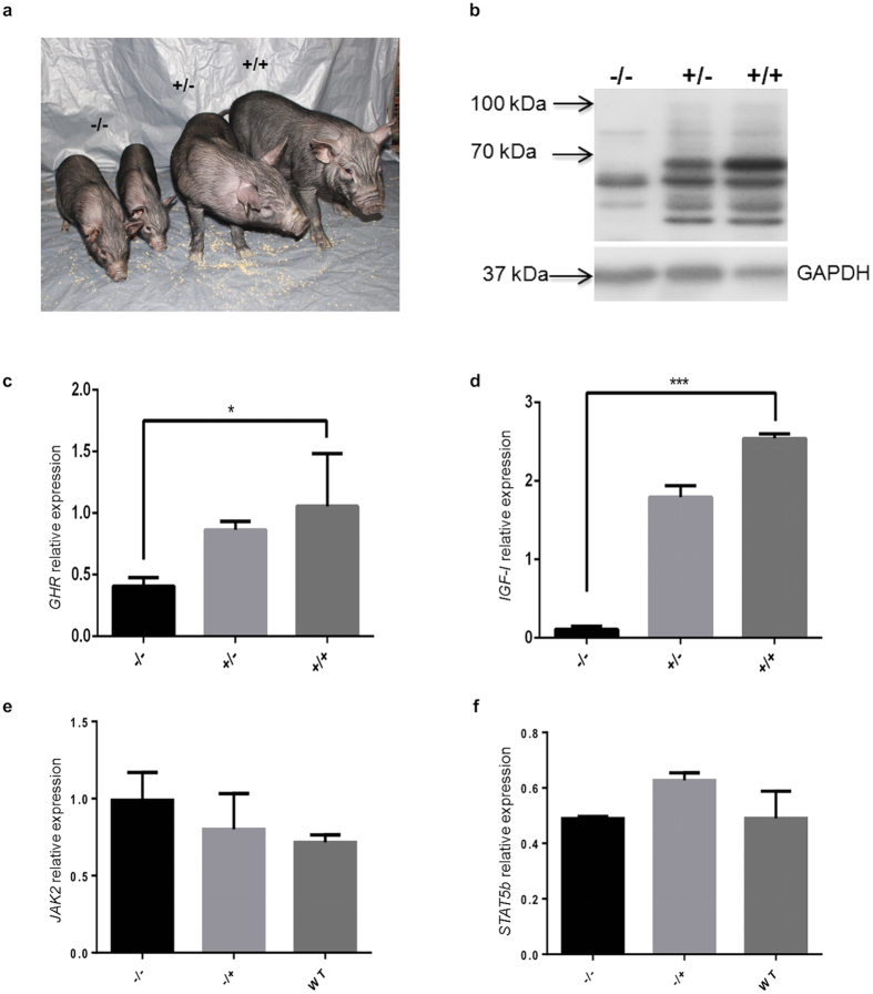
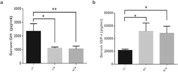
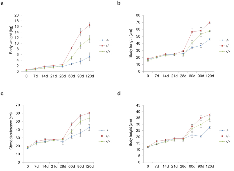
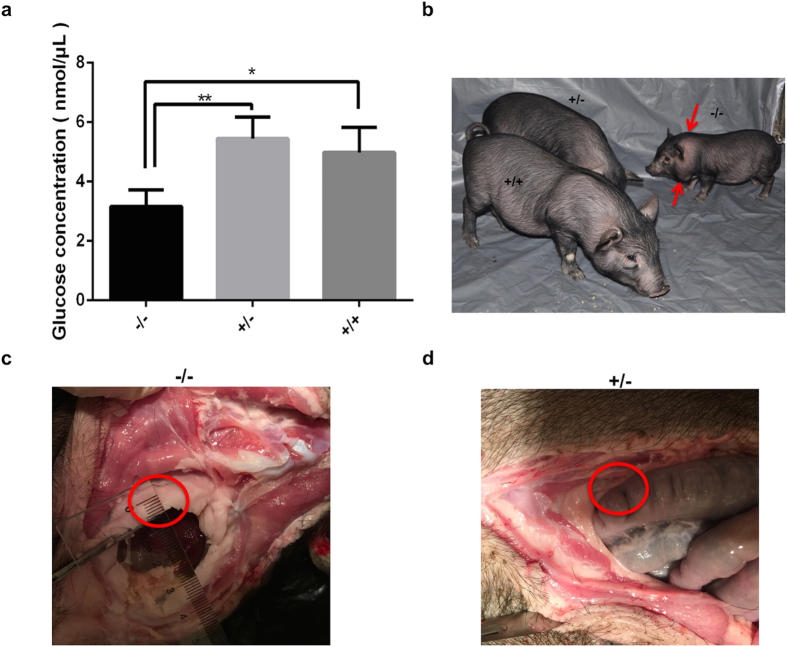
Laron syndrome is a rare disease caused by mutations of the growth hormone receptor (GHR), inheriting in an autosomal manner. To better understand the pathogenesis and to develop therapeutics, we generated a miniature pig model for this disease by employing ZFNs to knock out GHR gene. Three types of F0 heterozygous pigs (GHR(+/4bp), GHR(+/2bp), GHR(+/3bp)) were obtained and in which no significant phenotypes of Laron syndrome were observed. Prior to breed heterozygous pigs to homozygosity (GHR(4bp/4bp)), pig GHR transcript with the 4 bp insert was evaluated in vitro and was found to localize to the cytoplasm rather than the membrane. Moreover, this mutated transcript lost most of its signal transduction capability, although it could bind bGH. GHR(4bp/4bp) pigs showed a small body size and reduced body weight. Biochemically, these pigs exhibited significantly elevated levels of GH and decreased levels of IGF-I. These results resemble the phenotype observed in Laron patients, suggesting that these pigs could serve as an ideal model for Laron syndrome to bridge the gaps between mouse model and human.


Conflict of interest statement

The authors declare no competing financial interests.

Figures

Figure 1
Figure 1. Obtaining F0 heterozygous pigs through SCNT by knocking out GHR gene with ZFNs.
(a) Schematic representation of ZFNs targeting exon 6 of the pig GHR gene. E1–E10 represent the ten exons of the pig GHR gene, the lower-case characters and the upper-case characters are the spacers and the DNA binding sequences, respectively. (b) Identification of the cell clones for SCNT. The mutation types of GHR in different cell clones were small indels. (c) Identification of F0 heterozygous pigs by Sanger sequencing of PCR products. The results of 2 bp (TG) and 4 bp (TGGA) inserts are double peaks; WT indicates the result obtained from the wild-type pigs.
Figure 2
Figure 2. Evaluation of the pig GHR transcript with a 4 bp insert in vitro.
(a) RT-PCR of the 4 bp insert and the wild-type pig GHR transcripts in stably integrated DF1 cell clones. Pig-GHR+/+ and Pig-GHR4bp/4bp represent the wild-type and the 4 bp insert pig GHR, respectively; numbers represent the different cell clones obtained; WT represents the results obtained from un-transfected DF1 cells; P represents the plasmid control; and the sequencing results of the clones are also shown. (b) qRT-PCR of pig GHR mRNA expression in stably integrated DF1 cells. The expression levels were determined by the expression relative to GAPDH (an internal control). The data were combined from three independent experiments; the bars represent the means ± SD (n = 4–6 cell clones per group); **P < 0.01, *P < 0.05. (c) Western blot of pig GHR expression in stably integrated DF1 cells. The wild-type and mutated GHR are 100 kDa and 22 kDa, respectively; GAPDH was used as an internal control. (d) Determination of the localization of pig GHR in DF1 cells. Stably transfected DF1 cells were stained with the anti-Flag tag primary antibody (1:500) and Cy3-conjugated goat anti-mouse IgG antibody. DAPI (blue) staining indicates the nucleus. The data are representative of at least three independent experiments. Images were obtained with a 100x oil objective lens. (e) Dual-luciferase assay of the mutated pig GHR and the wild-type GHR. Dex represents Dexamethasone (200 nM); Spi+luciferase is the reporter vector used in this assay; GH (50 nM) was used to induce the signal transduction; the bars represent the means ± SD; *P < 0.05.
Figure 3
Figure 3. Knockout of the pig GHR gene in F2 homozygous pigs (GHR4bp/4bp refers to −/−).
(a) The −/− pigs have a smaller body size. (b) Western blot of GHR in F2 pigs. The −/−, +/− (GHR+/4bp) and +/+ (GHR+/+) represent different genotypes in the F2 generation. The expected sizes for pig GHR are 100 kDa (glycosylated) or 70 kDa (un-glycosylated); GAPDH was used as an internal control. The mRNA expression of GHR (c), IGF-I (d), JAK2 (e) and STAT5b (f) genes in the F2 generation was determined by qRT-PCR. The expression levels were determined by normalizing the expression relative to GAPDH. The data were combined from three independent experiments; the bars represent the means ± SD (at least three pigs per group); ***P < 0.001, *P < 0.05.
Figure 4
Figure 4. Growth retardation in the −/− pigs.
Determination of body weight (a), body length (b), chest circumference (c) and body height (d in the F2 generations suggests growth failure in the −/− pigs. The −/−, +/− (GHR+/4bp) and +/+ (GHR+/+) represent different genotypes in the F2 generation; the bars represent the means ± SD (at least three pigs per group); *P < 0.05.
Figure 5
Figure 5. Biochemical changes in the GH-IGF-I axis.
The −/− pigs showed significantly elevated level of GH (a) and reduced level of IGF-I (b). The −/−, +/− (GHR+/4bp) and +/+ (GHR+/+) represent different genotypes in the F2 generation; the bars represent the means ± SD (at least three pigs per group); **P < 0.01, *P < 0.05.
Figure 6
Figure 6. Glucose levels and obesity of the GHR homozygous KO pigs.
(a) The levels of glucose were determined by a Glucose Assay Kit (ab65333, Abcam). The −/−, +/− (GHR+/4bp) and +/+ (GHR+/+) represent different genotypes in the F2 generation, the bars represent the means ± SD (at least three pigs per group); **P < 0.01, *P < 0.05. (b) The −/−, +/− (GHR+/4bp) and +/+ (GHR+/+) pigs at age of two months old, red arrows showed the obesity observed in homozygous KO pigs. The thick layer of abdominal fat (as shown by red circles) was observed in −/− pig (c), while no such fat layer was observed in +/− (GHR+/4bp) pig (d). The pigs in (c) and (d) were three-and-half-month-old.

References

    1. Laron Z., Pertzelan A. & Mannheimer S. Genetic pituitary dwarfism with high serum concentation of growth hormone—a new inborn error of metabolism? Isr J Med Sci 2, 152–155 (1966). - PubMed
    1. Keret R., Pertzelan A., Zeharia A., Zadik Z. & Laron Z. Growth hormone (hGH) secretion and turnover in three patients with Laron-type dwarfism. Isr J Med Sci 24, 75–79 (1988). - PubMed
    1. Laron Z. et al.. Laron syndrome due to a post-receptor defect: response to IGF-I treatment. Isr J Med Sci 29, 757–763 (1993). - PubMed
    1. Godowski P. J. et al.. Characterization of the human growth hormone receptor gene and demonstration of a partial gene deletion in two patients with Laron-type dwarfism. Proceedings of the National Academy of Sciences of the United States of America 86, 8083–8087 (1989). - PMC - PubMed
    1. Bazan J. F. Structural Design and Molecular Evolution of a Cytokine Receptor Superfamily. Proceedings of the National Academy of Sciences of the United States of America 87, 6934–6938, doi: 10.1073/Pnas.87.18.6934 (1990). - DOI - PMC - PubMed

Publication types