Review
. 2015 Oct 28:9:248.
doi: 10.1186/s13256-015-0715-4.

Ultrasound plays a key role in imaging and management of genital angiomyofibroblastoma: a case report

Affiliations
Review

Ultrasound plays a key role in imaging and management of genital angiomyofibroblastoma: a case report

Benjamin Wolf et al. J Med Case Rep. .

Abstract

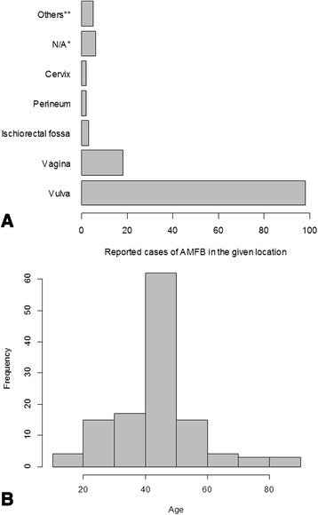
Introduction: Angiomyofibroblastoma is a benign, rare mesenchymal tumor arising from the genital tract of both men and women and was first described by Fletcher and colleagues in 1992. The tumor needs to be distinguished from other, similar lesions, such as deep and superficial aggressive angiomyxoma and cellular angiofibroma, because aggressive angiomyxoma demands much more extensive treatment. The vast majority of angiomyofibroblastomas arise from the vulva and appear as solid cystic masses on ultrasound images.

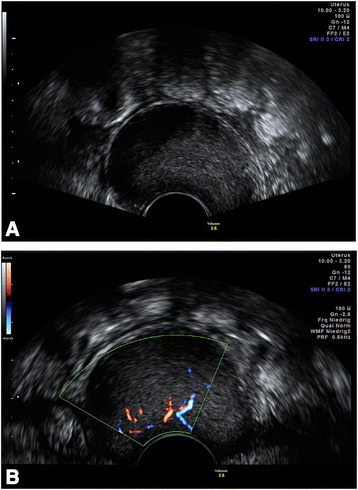
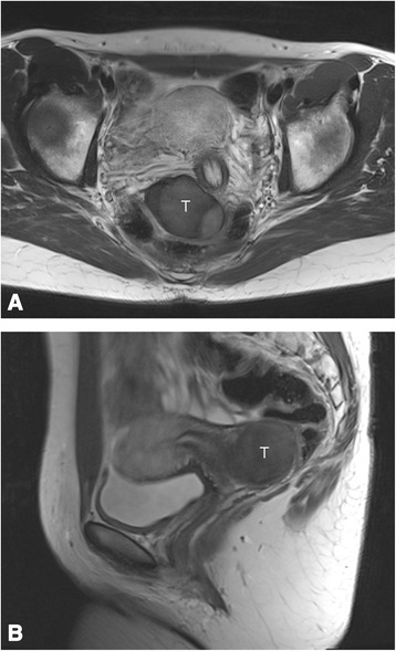
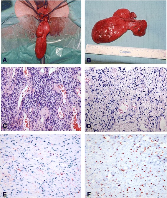
Case presentation: We report a case of a 35-year-old Caucasian woman with an angiomyofibroblastoma arising from the vagina. She presented with a painless mass of about 5cm in diameter that had a rather homogeneous, hypoechoic appearance on ultrasound images. The patient underwent surgical resection of the mass, which was subsequently diagnosed as angiomyofibroblastoma. We present sonographic and magnetic resonance imaging findings, intraoperative and histologic images, and a thorough review of the literature.

Conclusions: In our opinion, ultrasonography is the most valuable tool to establish a preoperative diagnosis of this tumor entity, differentiate it from other lesions of the female genital tract, and plan surgery accordingly. Even though it is a rare tumor, gynecologists should be able to recognize it and to differentiate it from other tumor entities that demand more aggressive treatment. We describe a different sonographic appearance of this tumor than previously reported.


Figures

Fig. 1
Fig. 1
a Transvaginal ultrasound image shows a well-demarcated, homogeneous mass of medium echogenicity (sagittal view). Intralesional septations can be seen only in Additional file 1. b Color Doppler imaging reveals intralesional vascularization
Fig. 2
Fig. 2
Coronal (a) and sagittal (b) T2-weighted magnetic resonance images obtained preoperatively show a homogeneous solid mass (T) displacing the cervix at left (seen best in coronal view)
Fig. 3
Fig. 3
a and b Intraoperative appearance of the tumor. The tumor was covered entirely with a frail membrane of pinkish gray appearance. c Cellular mesenchymal lesion with alternating cellularity intermingled with small blood vessels [hematoxylin and eosin (H&E) stain, original magnification ×109]. d Higher-magnification image representing thin-walled blood vessels surrounded by ovoid to spindle-shaped cells with some epithelioid appearance and abundant eosinophilic cytoplasm (H&E stain, original magnification ×241). Immunohistochemical staining for desmin with weak positivity. f Strong nuclear expression of estrogen receptor.
Fig. 4
Fig. 4
Analysis of all 125 cases of angiomyofibroblastoma in women reported to date (see also Additional file 2). a The number of cases of angiomyofibroblastoma occurring at each given location out of a total of 125 cases is shown. *N/A denotes that no information regarding the location was available. **Other locations include inguinal, urethral, fallopian tube, perianal, cul-de-sac, and retroperitoneal. b Histogram showing the age distribution of 116 patients with angiomyofibroblastoma (including men). For 21 patients, no age information was available for analysis. AMFB angiomyofibroblastoma

References

    1. Fletcher CD, Tsang WY, Fisher C, Lee KC, Chan JK. Angiomyofibroblastoma of the vulva: a benign neoplasm distinct from aggressive angiomyxoma. Am J Surg Pathol. 1992;16:373–82. doi: 10.1097/00000478-199204000-00006. - DOI - PubMed
    1. Poljak NK, Kljajić Z, Petricević J, Forempoher G, Simunić MM, Colović Z, et al. Polypoid angiomyofibroblastoma tumor of nasal cavity: case report. Coll Antropol. 2013;37:301–4. - PubMed
    1. Sutton BJ, Laudadio J. Aggressive angiomyxoma. Arch Pathol Lab Med. 2012;136:217–21. doi: 10.5858/arpa.2011-0056-RS. - DOI - PubMed
    1. Fetsch JF, Laskin WB, Lefkowitz M, Kindblom LG, Meis-Kindblom JM. Aggressive angiomyxoma: a clinicopathologic study of 29 female patients. Cancer. 1996;78:79–90. doi: 10.1002/(SICI)1097-0142(19960701)78:1<79::AID-CNCR13>3.0.CO;2-4. - DOI - PubMed
    1. Chan YM, Hon E, Ngai SW, Ng TY, Wong LC, Chan IM. Aggressive angiomyxoma in females: is radical resection the only option? Acta Obstet Gynecol Scand. 2000;79:216–20. doi: 10.1080/j.1600-0412.2000.079003216.x. - DOI - PubMed