. 2015 Oct 28;35(43):14457-66.
doi: 10.1523/JNEUROSCI.1451-15.2015.

Voltage-Mediated Control of Spontaneous Bundle Oscillations in Saccular Hair Cells

Affiliations

Voltage-Mediated Control of Spontaneous Bundle Oscillations in Saccular Hair Cells

Sebastiaan W F Meenderink et al. J Neurosci. .

Abstract

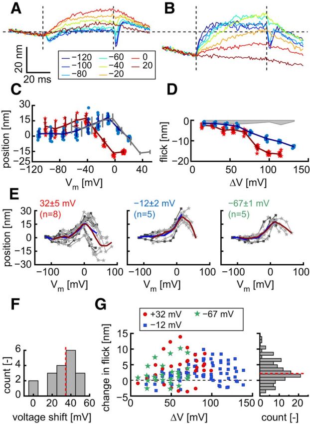
Hair cells of the vertebrate vestibular and auditory systems convert mechanical inputs into electrical signals that are relayed to the brain. This transduction involves mechanically gated ion channels that open following the deflection of mechanoreceptive hair bundles that reside on top of these cells. The mechano-electrical transduction includes one or more active feedback mechanisms to keep the mechanically gated ion channels in their most sensitive operating range. Coupling between the gating of the mechanosensitive ion channels and this adaptation mechanism leads to the occurrence of spontaneous limit-cycle oscillations, which indeed have been observed in vitro in hair cells from the frog sacculus and the turtle basilar papilla. We obtained simultaneous optical and electrophysiological recordings from bullfrog saccular hair cells with such spontaneously oscillating hair bundles. The spontaneous bundle oscillations allowed us to characterize several properties of mechano-electrical transduction without artificial loading the hair bundle with a mechanical stimulus probe. We show that the membrane potential of the hair cell can modulate or fully suppress innate oscillations, thus controlling the dynamic state of the bundle. We further demonstrate that this control is exerted by affecting the internal calcium concentration, which sets the resting open probability of the mechanosensitive channels. The auditory and vestibular systems could use the membrane potential of hair cells, possibly controlled via efferent innervation, to tune the dynamic states of the cells.

Keywords: frog; hair cell; sacculus; spontaneous oscillations.


Figures

Figure 1.
Figure 1.
Transduction currents are correlated with spontaneous hair bundle oscillations. A, Bundle position (black) and MET current (gray) for a single hair cell that was held at a membrane potential of −50 mV in LCP solution. Upward deflections indicate hair bundle motion toward the tallest row of stereovilli (open) and an inward MET current (in), respectively. This differs from convention, where outward current is plotted as positive; it aids in observing the correlation between bundle position and MET current. The hair bundle oscillated spontaneously at 42.4 Hz, and the two recordings were well correlated (r = 0.73). The hair bundle exhibited both full limit-cycle oscillations and motions of smaller amplitude (arrowheads) that likewise had a correlate in the MET current. B, Bundle position (black) and MET current (gray) for a hair cell (Vm = −50 mV) before, during, and after exposure of its apical surface to LCP containing 100 μm gentamicin. The presence of gentamicin, a blocker of the MET channels, abolishes both the spontaneous bundle oscillations and the measured currents. C, Bundle position (black) and MET current (gray) for a different hair cell that was held at a membrane potential of −75 mV. The hair bundle was in artificial endolymph that had ∼97% of the monovalent cations replaced with N-methyl-d-glucamine (Table 1). The spontaneous oscillations (at 15.5 Hz) did not have an obviously visible correlate in the recorded current (r = −0.38). D, Scatter plot of the bundle position versus the MET current for the recordings in C, partitioned in two clusters (different circles). Between these clusters, the mean inward current (white stars) was significantly larger when the bundle was deflected toward the tallest row of sterovilli (Welch's t test, t(3718) = −35.7, p < 0.001).
Figure 2.
Figure 2.
Membrane potential controls the occurrence of spontaneous hair bundle oscillations. A, Series of recordings of bundle position (black) and MET current (gray) for a single hair cell that was held at different membrane potentials (command voltages, in mV, are indicated on the right). Both bundle motion toward the tallest row of stereovilli (open) and the inward current (in) are positive. B–E, Various parameters as a function of Vm that were derived from recordings in 11 different hair cells. Each cell is represented by a series of circles, connected by lines. In all panels, each circle represents the mean result within an equal Vm segment (see Materials and Methods). The white line and shaded gray area represent the mean and SD across cells, respectively. Data were omitted if the bundle did not oscillate spontaneously. B, Fraction of time that the bundle is deflected toward the tallest row of sterovilli. C, Mean instantaneous frequency of spontaneous bundle oscillations. D, Amplitude of the bundle oscillation. E, Magnitude of the transduction current. All recordings were obtained from cells with their hair bundles exposed to LCP solution.
Figure 3.
Figure 3.
Gating force of the MET channels. A, Hair bundle position for a hair cell that was clamped to a membrane potential of −30 mV (gray line). Motion toward the tallest row of stereovilli is positive. Those sections of the curve that corresponded to the initial, fast motions toward (“up”; red circles) or away from the tallest row of stereovilli (“down”; blue stars) were selected to be fit with a single Boltzmann function (see Eq. 2). B, Scatter plot of instantaneous current versus displacement for the recording in A, including the fits that correspond to motions in the “up” (solid red line) and the “down” (solid blue line) directions. Dashed lines indicate extrapolations of the fits beyond the extent of bundle excursions. C, Comparison between Imax from the fit and the size of the transduction current (from Fig. 2E). Solid black line indicates the straight-line fit to these data (y = 1.07 (±0.06) x − 32 (±0.06); r2 = 0.86). Dashed line indicates equality. D, Similar comparison to C, obtained for the mean open probability of the MET complex (y = 1.02 (±0.05) x + 0.03 (±0.02); r2 = 0.89). Gating force (z) for motion toward (E) and away from (F) the tallest row of stereovilli. Error bars indicate ±1 SD.
Figure 4.
Figure 4.
Changes in the membrane potential elicit two distinct responses in the position of the hair bundle. A, Hair bundle position in response to a series of steps in the membrane potential. The two columns of traces are obtained from one cell, stepped from a membrane potential of 34 mV (left) and −68 mV (right). Top, Command voltage. Middle, Measured currents. Bottom, Bundle position. Both inward current (in) and motion toward the tallest row of stereovilli (open) are positive. Vertical dashed lines indicate the onset and offset of the voltage steps. Horizontal dashed lines indicate zero. For the position, this corresponds to zero position from B (for its calculation, see Materials and Methods). B, Mean (±SD) position for the recordings in A as a function of membrane potential. C, Mean (±SD) size of the flick that occurred immediately after membrane depolarizations for the recordings in A, as a function of the change in the membrane potential. Shaded gray area represents the noise floor for the estimate of the flick size. D, E, Similar to B, C, but show the mean (±SEM) for multiple hair cells (number of cells is given in parentheses in the legend).
Figure 5.
Figure 5.
Effect of the membrane potential on hair bundle position is calcium-mediated. A, Position of a hair bundle exposed to perilymph in response to a series of steps in the membrane potential (from 20 mV). See legend (in mV) for the step command voltages. B, Similar recording from the same hair bundle as in A, exposed to LCP solution. Because of increased currents, voltage steps are from 12 mV. Vertical dashed lines indicate the onset and offset of the steps. Horizontal dashed line indicates zero bundle position from C. Scale bars apply to A, B. C, Mean (±SD) hair bundle position and individual responses in perilymph (red stars), and LCP solutions (blue circles), for the recordings in A, B. Gray curve represents the data obtained in LCP solution but shifted horizontally to maximally overlap with the data obtained in perilymph (shift = 38.4 mV). D, Size of the flick for the recordings in A, B, same symbols as in C. Shaded gray area represents the noise floor for the estimate of the flick size. E, Mean bundle positions in perilymph (dark gray squares) and horizontally shifted mean bundle positions in LCP (light gray stars) for recordings stepped from different membrane potentials. Blue and red lines indicate loess trend lines (second-order polynomial; bandwidth: 35% of data; tri-cube weight function) for the perilymph and LCP data, respectively. F, Histogram (10 mV bins) of the applied horizontal shifts in E to maximally overlap the bundle positions. The median shift of 34.5 mV (dashed red line; interquartile range: 24.6 to 40.1 mV) matched the change in the Nernst potential for calcium (34.8 mV) between the two solutions. G, Left, Pairwise comparison between the flick size in perilymph and LCP. Positive values indicate that the flick is larger in LCP. Right, Histogram (1 nm bins) of these data, which had a median value of 2.7 nm (red dashed line) that was different from zero (Wilcoxon signed rank; Z = −7.29, p < 0.001).

References

    1. Assad JA, Hacohen N, Corey DP. Voltage dependence of adaptation and active bundle movement of bullfrog saccular hair cells. Proc Natl Acad Soc U S A. 1989;86:2918–2922. doi: 10.1073/pnas.86.8.2918. - DOI - PMC - PubMed
    1. Benser ME, Marquis RE, Hudspeth AJ. Rapid, active hair bundle movements in hair cells from the bullfrog's sacculus. J Neurosci. 1996;16:5629–5643. - PMC - PubMed
    1. Beurg M, Tan X, Fettiplace R. A prestin motor in chicken auditory hair cells: active force generation in a nonmammalian species. Neuron. 2013;79:69–81. doi: 10.1016/j.neuron.2013.05.018. - DOI - PMC - PubMed
    1. Bozovic D, Hudspeth AJ. Hair-bundle movements elicited by transepithelial electrical stimulation of hair cells in the sacculus of the bullfrog. Proc Natl Acad Soc U S A. 2003;100:958–963. doi: 10.1073/pnas.0337433100. - DOI - PMC - PubMed
    1. Camalet S, Duke T, Jülicher F, Prost J. Auditory sensitivity provided by self-tuned critical oscillations of hair cells. Proc Natl Acad Soc U S A. 2000;97:3183–3188. doi: 10.1073/pnas.97.7.3183. - DOI - PMC - PubMed

Publication types

MeSH terms