. 2015 Oct 28;35(43):14533-43.
doi: 10.1523/JNEUROSCI.1776-15.2015.

Expression of ESR1 in Glutamatergic and GABAergic Neurons Is Essential for Normal Puberty Onset, Estrogen Feedback, and Fertility in Female Mice

Affiliations

Expression of ESR1 in Glutamatergic and GABAergic Neurons Is Essential for Normal Puberty Onset, Estrogen Feedback, and Fertility in Female Mice

Rachel Y Cheong et al. J Neurosci. .

Abstract

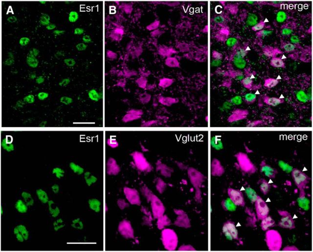
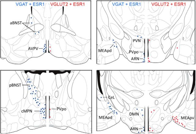
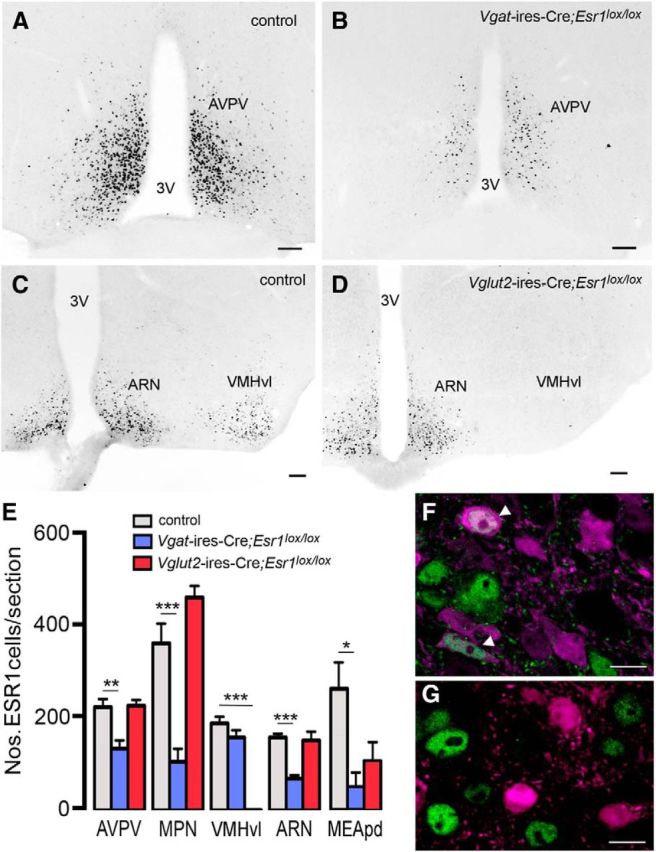
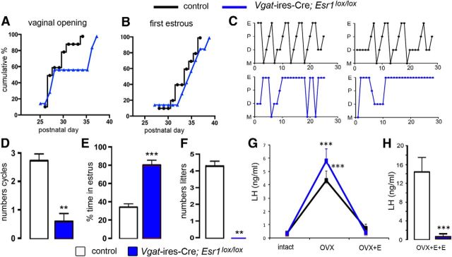
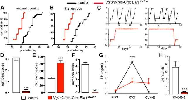
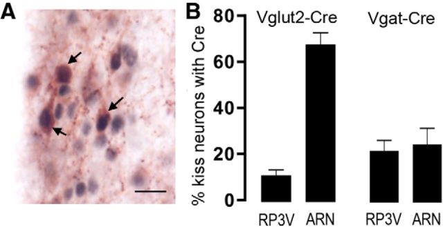
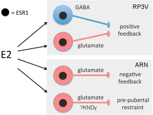
Circulating estradiol exerts a profound influence on the activity of the gonadotropin-releasing hormone (GnRH) neuronal network controlling fertility. Using genetic strategies enabling neuron-specific deletion of estrogen receptor α (Esr1), we examine here whether estradiol-modulated GABA and glutamate transmission are critical for the functioning of the GnRH neuron network in the female mouse. Using Vgat- and Vglut2-ires-Cre knock-in mice and ESR1 immunohistochemistry, we demonstrate that subpopulations of GABA and glutamate neurons throughout the limbic forebrain express ESR1, with ESR1-GABAergic neurons being more widespread and numerous than ESR1-glutamatergic neurons. We crossed Vgat- and Vglut2-ires-Cre mice with an Esr1(lox/lox) line to generate animals with GABA-neuron-specific or glutamate-neuron-specific deletion of Esr1. Vgat-ires-Cre;Esr1(lox/lox) mice were infertile, with abnormal estrous cycles, and exhibited a complete failure of the estrogen positive feedback mechanism responsible for the preovulatory GnRH surge. However, puberty onset and estrogen negative feedback were normal. Vglut2-ires-Cre;Esr1(lox/lox) mice were also infertile but displayed a wider range of deficits, including advanced puberty onset, abnormal negative feedback, and abolished positive feedback. Whereas <25% of preoptic kisspeptin neurons expressed Cre in Vgat- and Vglut2-ires-Cre lines, ∼70% of arcuate kisspeptin neurons were targeted in Vglut2-ires-Cre;Esr1(lox/lox) mice, possibly contributing to their advanced puberty phenotype. These observations show that, unexpectedly, ESR1-GABA neurons are only essential for the positive feedback mechanism. In contrast, we reveal the key importance of ESR1 in glutamatergic neurons for multiple estrogen feedback loops within the GnRH neuronal network required for fertility in the female mouse.

Keywords: GABA; GnRH; estradiol; glutamate.


Figures

Figure 1.
Figure 1.
ESR1-expression in GABA and VGLUT2 neurons in the female mouse brain. A–F, Dual-label immunohistochemistry for ESR1 (Alexa Fluor 488 green nuclei in A, D) and VGAT (tdTomato pseudocoloured as magenta, B) or VGLUT2 (E) from the AVPV and ARN, respectively, of Vgat- and Vglut2-ires-Cre mice crossed with a tdTomato reporter. Merged images show individual AVPV VGAT (C) or ARN VGLUT2 (F) neurons expressing ESR1 (white nuclei, arrowheads). Scale bars, 10 μm.
Figure 2.
Figure 2.
Distribution and density of ESR1-expressing GABA and VGLUT2 neurons in the female mouse brain. Schematic diagrams adapted from Franklin and Paxinos (1997) showing the topography of VGAT-ESR1 (blue) and VGLUT2-ESR1 (red) neurons at four different levels through the female mouse forebrain. Each dot represents 10 dual-labeled cells in a 30-μm-thick coronal brain section. aBNST, anterior bed nucleus of the stria terminalis; CeL, lateral central amygdala; DMN, dorsomedial nucleus; MEApd, posterior dorsal medial amygdala; MEApv, posterior ventral medial amygdala; pBNST, posterior bed nucleus of the stria terminalis; PVpo, preoptic periventricular nucleus; PVN, paraventricular nucleus.
Figure 3.
Figure 3.
ESR1-expression in Vgat-ires-Cre;Esr1lox/lox and Vglut2-ires-Cre;Esr1lox/lox mice. A, B, ESR1-immunoreactive nuclei in the AVPV of a female control Vgat-ires-Cre mouse (A) and mutant Vgat-ires-Cre;Esr1lox/lox mouse (B). C, D, ESR1-immunoreactive nuclei in the mediobasal hypothalamus of a female control Vglut2-ires-Cre mouse (C) and mutant Vglut2-ires-Cre;Esr1lox/lox mouse (D). 3V, Third ventricle. Scale bars, 70 μm. E, Histograms showing mean ± SEM numbers of ESR1 cells per section in control (n = 16), Vgat-ires-Cre;Esr1lox/lox (n = 7), and Vglut2-ires-Cre;Esr1lox/lox (n = 5) mice in 5 different brain regions. **p < 0.01, ***p < 0.001 (ANOVA with post hoc Tukey multiple comparison tests). F, G, ESR1 is present in VGLUT2 neurons in control mice (arrowheads, F) but deleted from ARN VGLUT2 neurons in Vglut2-ires-Cre;Esr1lox/lox;tdTomato mice (G). Scale bar, 5 μm.
Figure 4.
Figure 4.
Reproductive profile of Vgat-ires-Cre;Esr1lox/lox mice. A, B, Puberty onset for control (black, n = 16) and mutant (blue, n = 7) mice displayed as cumulative percentage charts for vaginal opening (A) and first estrous (B). C, Representative adult estrous cycle profiles for two control (top) and two mutant (below) mice. D, E, Histograms showing the mean ± SEM number of complete estrous cycles (D) and percentage time spent in estrous (E) in control (n = 20) and mutant Vgat-ires-Cre;Esr1lox/lox (n = 9) mice. **p < 0.01, ***p < 0.001 (Mann–Whitney U tests). F, Histogram showing the mean ± SEM number of litters born to control (n = 5) and mutant (n = 5) female mice over a 3 month mating period. **p < 0.01 (Mann–Whitney U tests). G, Estrogen negative feedback in control (black, n = 7) and mutant (blue, n = 6) mice showing mean ± SEM changes in LH secretion after OVX and OVX+E. ***p < 0.01 versus intact and OVX+E (repeated-measures ANOVA with Bonferroni post hoc tests). H, Positive feedback mean ± SEM levels of LH in ovariectomized control (n = 7) and mutant (n = 6) mice treated with estradiol (OVX+E+E) to evoke the GnRH/LH surge. ***p < 0.001 (Mann–Whitney U test).
Figure 5.
Figure 5.
Reproductive profile of Vglut2-ires-Cre;Esr1lox/lox mice. A, B, Puberty onset for control (black, n = 16) and mutant (red, n = 8) mice displayed as cumulative percentage charts for vaginal opening (A) and first estrous (B). C, Representative adult estrous cycle profiles for two control (top) and two mutant (below) mice. D, E, Histograms showing the mean ± SEM number of complete estrous cycles (D) and percentage time spent in estrous (E) in control (n = 20) and mutant Vglut2-ires-Cre;Esr1lox/lox (n = 7) mice. ***p < 0.001 (Mann–Whitney U tests). F, Histogram showing the mean ± SEM number of litters born to control (n = 5) and mutant (n = 5) female mice over a 3 month mating period. **p < 0.01 (Mann–Whitney U tests). G, Estrogen negative feedback in control (black, n = 7) and mutant (red, n = 5) mice showing mean ± SEM changes in LH secretion after OVX and OVX+E. ***p < 0.01 versus intact and OVX+E (repeated-measures ANOVA with Bonferroni post hoc tests). H, Positive feedback mean ± SEM levels of LH in ovariectomized control (n = 8) and mutant (n = 5) mice treated with estradiol (OVX+E+E) to evoke the GnRH/LH surge. ***p < 0.001 (Mann–Whitney U test).
Figure 6.
Figure 6.
Expression of Cre in kisspeptin neurons of Vgat- and Vglut2-Cre mouse lines. A, Dual-label immunohistochemistry for Cre (black nuclei) and kisspeptin (brown cytoplasm) in the AVPV of a Vglut2-ires-Cre mouse. Three dual-labeled cells are indicated with arrows. Scale bar, 20 μm. B, Histograms showing the mean ± SEM percentage of kisspeptin neurons expressing Cre in the RP3V and ARN of Vglut2-ires-Cre (n = 4) and Vgat-ires-Cre (n = 4) adult female diestrous mice.
Figure 7.
Figure 7.
Schematic diagram summarizing the likely essential roles of ESR1-expressing GABA (blue) and glutamate (red) neurons in the various estrogen feedback mechanisms within the GnRH neuronal network. The possible locations of the cell bodies of the respective GABA and glutamate inputs within each feedback loop are indicated within each gray box. KNDy, Kisspeptin-Neurokinin B-Dynoprhin neuron.

References

    1. Adachi S, Yamada S, Takatsu Y, Matsui H, Kinoshita M, Takase K, Sugiura H, Ohtaki T, Matsumoto H, Uenoyama Y, Tsukamura H, Inoue K, Maeda K. Involvement of anteroventral periventricular metastin/kisspeptin neurons in estrogen positive feedback action on luteinizing hormone release in female rats. J Reprod Dev. 2007;53:367–378. doi: 10.1262/jrd.18146. - DOI - PubMed
    1. Brann DW. Glutamate: a major excitatory transmitter in neuroendocrine regulation. Neuroendocrinology. 1995;61:213–225. doi: 10.1159/000126843. - DOI - PubMed
    1. Brann DW, Mahesh VB. Endogenous excitatory amino acid involvement in the preovulatory and steroid-induced surge of gonadotropins in the female rat. Endocrinology. 1991;128:1541–1547. doi: 10.1210/endo-128-3-1541. - DOI - PubMed
    1. Campos P, Herbison AE. Optogenetic activation of GnRH neurons reveals minimal requirements for pulsatile luteinizing hormone secretion. Proc Natl Acad Sci U S A. 2014;111:18387–18392. doi: 10.1073/pnas.1415226112. - DOI - PMC - PubMed
    1. Casanova E, Fehsenfeld S, Mantamadiotis T, Lemberger T, Greiner E, Stewart AF, Schütz G. A CamKIIalpha iCre BAC allows brain-specific gene inactivation. Genesis. 2001;31:37–42. doi: 10.1002/gene.1078. - DOI - PubMed

Publication types

MeSH terms