. 2015 Oct 28;7(311):311ra173.
doi: 10.1126/scitranslmed.aac4269.

Inflammation activation and resolution in human tendon disease

Affiliations

Inflammation activation and resolution in human tendon disease

Stephanie G Dakin et al. Sci Transl Med. .

Abstract

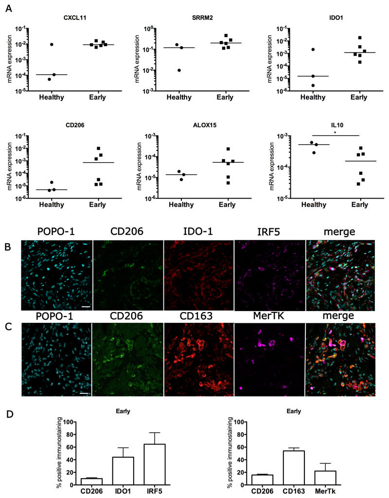
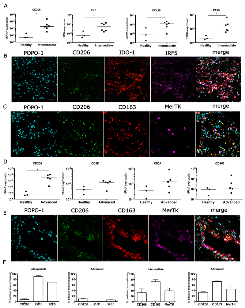
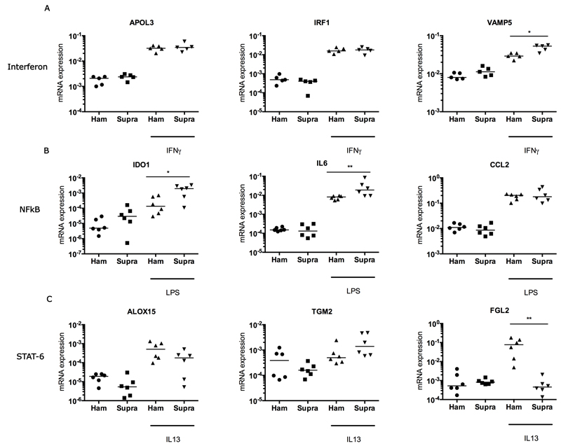
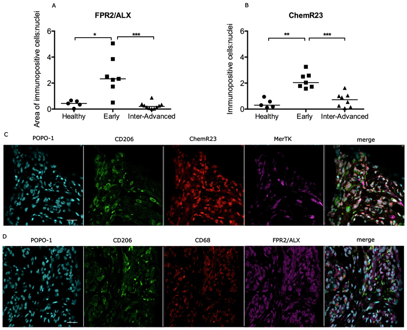
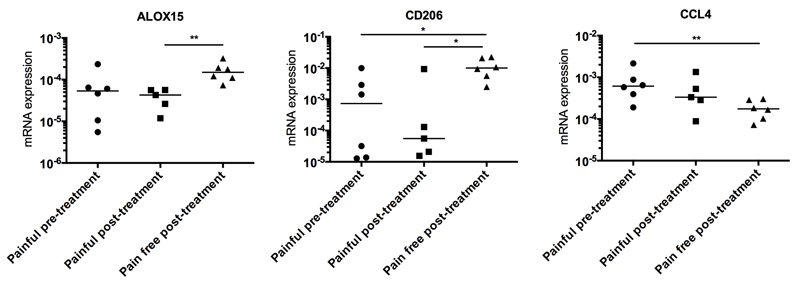
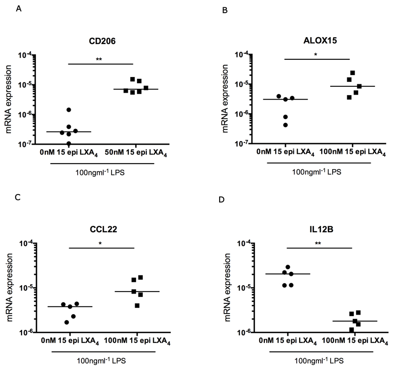
Improved understanding of the role of inflammation in tendon disease is required to facilitate therapeutic target discovery. We studied supraspinatus tendons from patients experiencing pain before and after surgical subacromial decompression treatment. Tendons were classified as having early, intermediate, or advanced disease, and inflammation was characterized through activation of pathways mediated by interferon (IFN), nuclear factor κB (NF-κB), glucocorticoid receptor, and signal transducer and activator of transcription 6 (STAT-6). Inflammation signatures revealed expression of genes and proteins induced by IFN and NF-κB in early-stage disease and genes and proteins induced by STAT-6 and glucocorticoid receptor activation in advanced-stage disease. The proresolving proteins FPR2/ALX and ChemR23 were increased in early-stage disease compared to intermediate- to advanced-stage disease. Patients who were pain-free after treatment had tendons with increased expression of CD206 and ALOX15 mRNA compared to tendons from patients who continued to experience pain after treatment, suggesting that these genes and their pathways may moderate tendon pain. Stromal cells from diseased tendons cultured in vitro showed increased expression of NF-κB and IFN target genes after treatment with lipopolysaccharide or IFNγ compared to stromal cells derived from healthy tendons. We identified 15-epi lipoxin A4, a stable lipoxin isoform derived from aspirin treatment, as potentially beneficial in the resolution of tendon inflammation.


Conflict of interest statement

Competing interests: The authors declare that they have no competing interests.

Figures

Figure 1
Figure 1
Immunohistochemistry showing CD14+ and CD68+ monocytes/macrophages in healthy and diseased human supraspinatus tendons. (A, B) Graphs show quantitative analysis of CD14+ cells (monocytes) and CD68+ cells (macrophages) in healthy tendons, and in early and intermediate-advanced stage diseased tendons. Bar shows median values. Statistically significant differences were calculated using Kruskal-Wallis with pairwise post-hoc Mann Whitney U tests. **p<0.01, ***p<0.001. Panel shows representative images of 3,3’-Diaminobenzidine immunostaining (brown) for CD14 and CD68 in healthy (C,F), early (D,G) and intermediate-advanced stages of tendon disease (E,H). Nuclear counterstain is haematoxylin. Scale bar=50µm. (I,J) Representative confocal immunofluorescence images showing dual labelling for CD14 (purple) and CD68 (green) in sections of early and advanced stage diseased tendons. Cyan represents POPO-1 nuclear counterstain. Scale bar = 20µm.
Figure 2
Figure 2
Expression of inflammation activation pathway genes and proteins in early stage diseased supraspinatus tendons compared to healthy subscapularis tendons. (A) A mixed gene expression signature was present in pre-treatment tendon samples with early disease (n=6) compared to healthy subscapularis control tendons (n=3). Shown is expression of genes induced by IFN including CXCL11, and Serine/arginine repetitive matrix 2 (SRRM2); induced by NF-κB including Indoleamine 2,3-dioxygenase 1 (IDO1) and induced by STAT-6 ALOX15 and CD206. Fold changes in gene expression are shown in Table S2. There was greater expression of IFN-induced genes including CXCL11 and SRRM2, and the NF-κB-induced gene IDO1 in early stage disease supraspinatus tendons compared to healthy subscapularis control tendons. Gene expression is normalized to β-actin; bar shows median value. (B, C) Representative immunofluorescence images of sections of pre-treatment early stage disease tendons stained for inflammation activation markers including those in the STAT-6 pathway (CD206, green), the glucocorticoid receptor pathway (CD163 red), the IFN pathway (IRF5, purple) and the NF-κB pathway (IDO-1, red). MerTK (purple) represents Mer tyrosine kinase, a tissue resident macrophage marker. Cyan represents POPO-1 nuclear counterstain. Scale bar = 20µm. (D) Quantitative analysis showing percentage of immunopositive staining for inflammation activation markers. Data are shown as mean and SEM.
Figure 3
Figure 3
Inflammation activation pathway signatures in intermediate and advanced stage disease tendons. (A) A mixed inflammation activation gene signature in intermediate stage diseased supraspinatus tendon samples (n=6) compared to healthy subscapularis tendons (n=3). (B, C) Representative immunofluorescence images of the inflammation activation protein signature in intermediate stage diseased tendons. (B) Markers indicate activation of the following pathways: STAT-6 (CD206, green), NF-κB (IDO-1, red), and IFN (IRF5, purple). (C) Markers indicate activation of the glucocorticoid receptor pathway (CD163, red); also shown is the tissue resident macrophage marker Mer tyrosine kinase (MerTK, purple). (D) A STAT-6/glucocorticoid receptor gene signature predominates in advanced stage diseased supraspinatus tendons (n=5) compared to healthy subscapularis tendons (n=3). Gene expression is normalized to β-actin. Statistically significant differences were calculated using pairwise Mann Whitney U tests. Bar represents median, *denotes p<0.05. (E) Representative immunofluorescence images of the inflammation activation protein signature in advanced stage diseased supraspinatus tendon. Shown is staining for the STAT-6 (CD206, green) and glucocorticoid receptor (CD163, red) activation pathways; tissue resident macrophages are stained with Mer tyrosine kinase (MerTK, purple). Cyan represents POPO-1 nuclear counterstain. Scale bar = 20µm. (F) Quantitative analysis showing the percentage of cells staining immunopositive for inflammation pathway proteins. Data are shown as mean and SEM.
Figure 4
Figure 4
Gene expression after treatment of cultured tendon-derived stromal cells with IFNγ, LPS or IL13 for 96 hours. Tendon stromal cells were derived from healthy hamstring or diseased supraspinatus tendons (n=6 donors for LPS and IL13 treatment, n=5 donors for IFNγ treatment). (A) Treatment of cells from healthy hamstring (Ham) or diseased supraspinatus tendons (Supra) with 20 ngmL-1 IFNγ. (B) Stimulation of cells from healthy hamstring (Ham) or diseased supraspinatus tendons (Supra) with 100 ngmL-1 LPS. (C) Stimulation of cells from healthy hamstring (Ham) or diseased supraspinatus tendons (Supra) with 20ngmL-1 IL13. Statistically significant differences were calculated using pairwise Mann Whitney U tests. Gene expression is normalized to β-actin; bar represents median. *p<0.05, **p<0.01.
Figure 5
Figure 5
Expression of pro-resolving proteins in healthy and diseased supraspinatus tendons. Quantitative analyses show increased expression of FPR2/ALX (A) and ChemR23 (B) in early stage disease tendons compared to intermediate-advanced stage disease tendons or healthy tendons. Bar represents median. Statistically significant differences were calculated using Kruskal-Wallis with pairwise post-hoc Mann Whitney U tests. *p<0.05, **p<0.01, ***p<0.001. Representative confocal immunofluorescence images showing staining for macrophages (CD206+, CD68+, MerTK+) and the pro-resolving proteins ChemR23 (red, C) and FPR2/ALX (purple, D) in sections of early stage tendon disease. Cyan represents nuclear counterstain. Scale bar = 20µm.
Figure 6
Figure 6
Expression of ALOX15, CD206 and CCL4 mRNA in biopsies from patients with early stage tendon disease before and after surgical sub-acromial decompression treatment. Pre-treatment tendinopathy samples from patients experiencing pain (n=6) were obtained after presentation to a referral clinic and prior to surgery. Biopsies were also collected from patients 2 to 4 years post-treatment. Post-treatment patients either had persistent pain (n=5) or were pain-free (n=6).Statistically significant differences were calculated using pairwise Mann Whitney U tests. Gene expression is normalized to β-actin; bar represents median. * p<0.05, **p<0.01.
Figure 7
Figure 7
The aspirin metabolite 15-epi LXA4 modulates inflammation in LPS-treated stromal cells derived from diseased tendons. Tendon cells were derived from intermediate stage disease supraspinatus tendons from a minimum of 5 patients. (A) Tendon-derived cells were treated with 100ngmL-1 LPS in medium containing 50nM 15-epi LXA4 for 96 hours. There was increased expression of CD206 mRNA after treatment with LPS and 15-epi LXA4 compared to LPS-alone (cells derived from tendons from 6 patients). (B,C,D) Tendon cells were treated with 100ngmL-1 LPS in medium containing 100nM 15-epi LXA4 for 96 hours. There was increased expression of ALOX15 mRNA (B) and CCL22 mRNA (C), and reduced expression of IL12B mRNA (D) compared to LPS-alone (cells derived from tendons from 5 patients). Statistically significant differences were calculated using pairwise Mann Whitney U tests. Gene expression is normalized to β-actin. Bar shows median values. * p<0.05, **p<0.01.

References

    1. Hinsley H, Nicholls A, Daines M, Wallace G, Arden N, Carr A. Classification of rotator cuff tendinopathy using high definition ultrasound. Muscles ligaments and tendons journal. 2014;4:391–397. - PMC - PubMed
    1. Murphy RJ, Carr AJ. Shoulder pain. BMJ clinical evidence. 2010;2010 - PMC - PubMed
    1. Kannus P, Jozsa L. Histopathological changes preceding spontaneous rupture of a tendon. A controlled study of 891 patients. J Bone Joint Surg Am. 1991;73:1507–1525. - PubMed
    1. Dakin SG, Smith RK, Heinegard D, Onnerfjord P, Khabut A, Dudhia J. Proteomic analysis of tendon extracellular matrix reveals disease stage-specific fragmentation and differential cleavage of COMP (cartilage oligomeric matrix protein) J Biol Chem. 2014;289:4919–4927. - PMC - PubMed
    1. Judge A, Murphy RJ, Maxwell R, Arden NK, Carr AJ. Temporal trends and geographical variation in the use of subacromial decompression and rotator cuff repair of the shoulder in England. The bone & joint journal. 2014;96-B:70–74. - PubMed

Publication types

MeSH terms

LinkOut - more resources