. 2016 Apr;65(4):575-83.
doi: 10.1136/gutjnl-2015-309728. Epub 2015 Oct 28.

The gut microbiota plays a protective role in the host defence against pneumococcal pneumonia

Affiliations

The gut microbiota plays a protective role in the host defence against pneumococcal pneumonia

Tim J Schuijt et al. Gut. 2016 Apr.

Abstract

Objective: Pneumonia accounts for more deaths than any other infectious disease worldwide. The intestinal microbiota supports local mucosal immunity and is increasingly recognised as an important modulator of the systemic immune system. The precise role of the gut microbiota in bacterial pneumonia, however, is unknown. Here, we investigate the function of the gut microbiota in the host defence against Streptococcus pneumoniae infections.

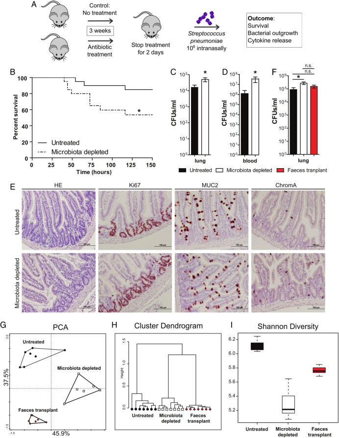
Design: We depleted the gut microbiota in C57BL/6 mice and subsequently infected them intranasally with S. pneumoniae. We then performed survival and faecal microbiota transplantation (FMT) experiments and measured parameters of inflammation and alveolar macrophage whole-genome responses.

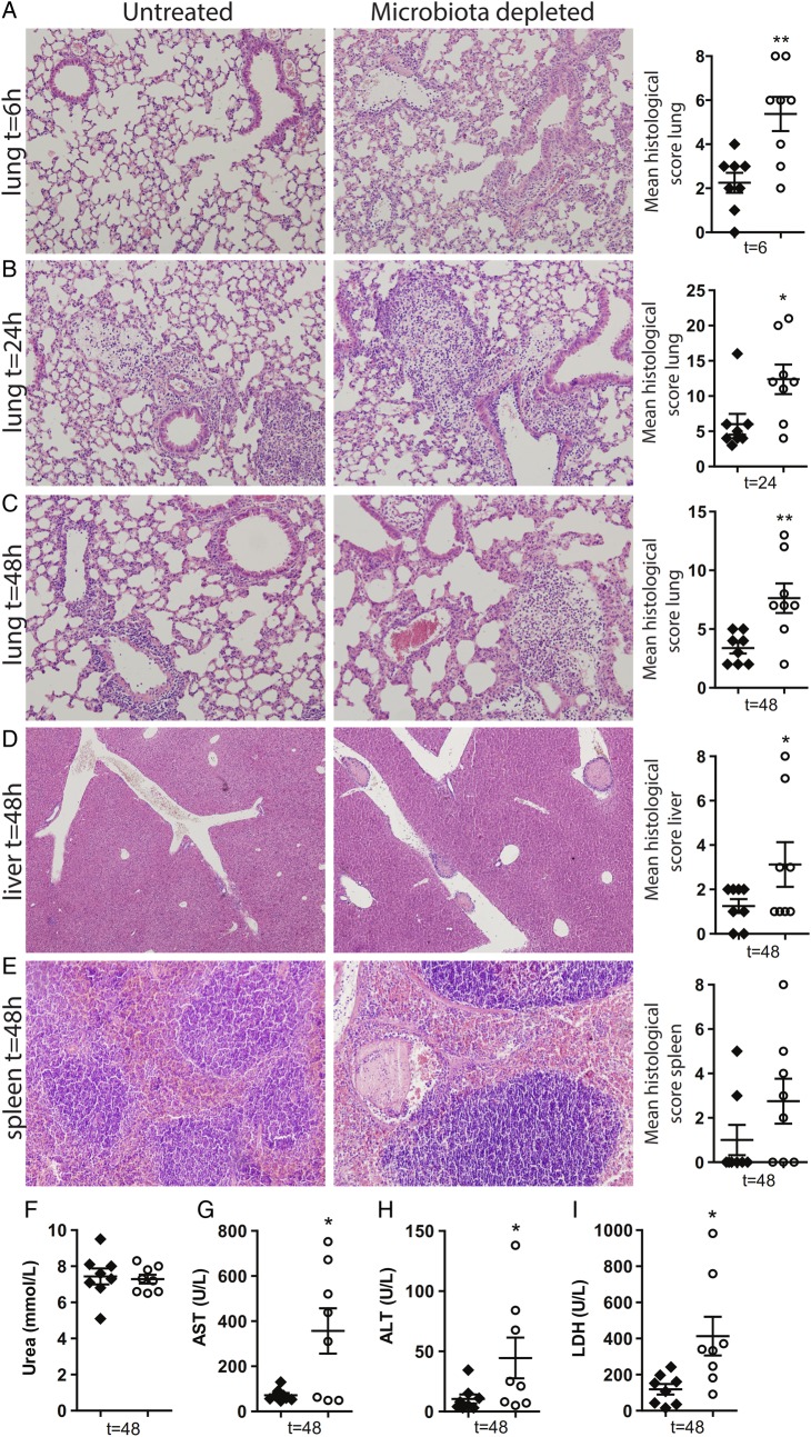
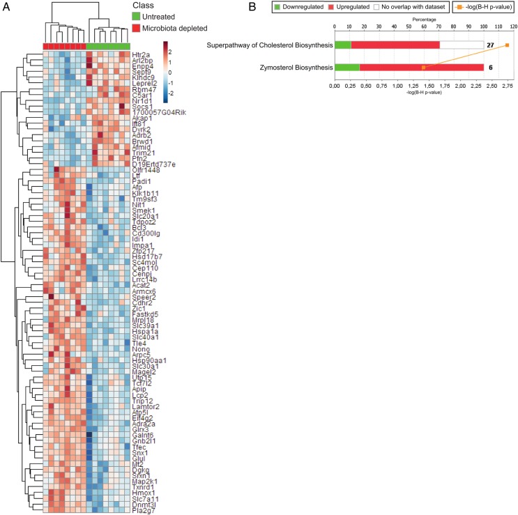
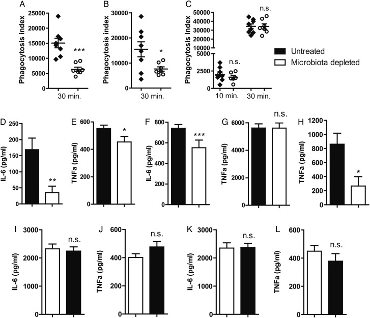
Results: We found that the gut microbiota protects the host during pneumococcal pneumonia, as reflected by increased bacterial dissemination, inflammation, organ damage and mortality in microbiota-depleted mice compared with controls. FMT in gut microbiota-depleted mice led to a normalisation of pulmonary bacterial counts and tumour necrosis factor-α and interleukin-10 levels 6 h after pneumococcal infection. Whole-genome mapping of alveolar macrophages showed upregulation of metabolic pathways in the absence of a healthy gut microbiota. This upregulation correlated with an altered cellular responsiveness, reflected by a reduced responsiveness to lipopolysaccharide and lipoteichoic acid. Compared with controls, alveolar macrophages derived from gut microbiota-depleted mice showed a diminished capacity to phagocytose S. pneumoniae.

Conclusions: This study identifies the intestinal microbiota as a protective mediator during pneumococcal pneumonia. The gut microbiota enhances primary alveolar macrophage function. Novel therapeutic strategies could exploit the gut-lung axis in bacterial infections.

Keywords: BACTERIAL INFECTION; BACTERIAL PATHOGENESIS; IMMUNOLOGY; INTESTINAL MICROBIOLOGY; SEPSIS.


Figures

Figure 1
Figure 1
Protective role of the gut microbiota during pneumococcal pneumonia. (A) Experimental design. Group of eight wild-type mice were treated for 3 weeks with broad-spectrum antibiotics (ampicillin, neomycin, metronidazole and vancomycin) in their drinking water compared with untreated controls. Two days post treatment mice received an intranasal challenge with 1×10⁶ colony forming units (CFU) of Streptococcus pneumoniae. Subsequently, mouse survival, bacterial outgrowth and cytokine release were determined at various time points post S. pneumoniae infection. (B) Survival of mice treated with broad-spectrum antibiotics compared with untreated controls before intranasal challenge with 1×10⁶ CFU of S. pneumoniae. (C) Pulmonary bacterial counts 6 h after S. pneumoniae infection in untreated (black) and microbiota-depleted (white) mice. (D) Blood bacterial counts 48 h after S. pneumoniae infection in untreated (black) and microbiota-depleted (white) mice. (E) Representative small intestine sections of untreated (upper row) and microbiota-depleted (lower row) mice demonstrate intact epithelial integrity with H&E, Ki67 (proliferation restricted in the crypt), MUC2 (goblet cell differentiation) and ChromograninA (neuroendocrine cell differentiation) stainings in both groups. (F) Effect of faecal microbiota transplantation to gut microbiota-depleted mice on lung bacterial counts 6 h after intranasal S. pneumoniae infection. (G) The magnitude by which the antibiotic protocol depleted the gut microbiota was assessed using a phylogenetic microarray in which microbiota composition samples were clustered based on principal component analysis (PCA), (H) Pearson Clustering and (I) the Shannon Diversity Index. Group size is 8–12 per group; results are shown as means±SEM; n.s. denotes not significant; *p<0.05.
Figure 2
Figure 2
The gut microbiota protects against organ failure during Streptococcus pneumoniae-induced sepsis. (A–C) Representative lung slides of untreated (left) and microbiota-depleted (right) mice infected with 1×10⁶ colony forming units (CFU) of S. pneumoniae via the intranasal route and euthanised at indicated time points (6, 24 and 48 h) thereafter to assess pulmonary inflammation and total lung histopathology scores (see Materials and methods). H&E staining; original magnification, ×100. (D and E) Liver and spleen histology shown 48 h after infection. H&E staining; original magnification, ×200 for liver, ×100 for spleen. (F) Systemic blood urea nitrogen (BUN), (G) aspartate aminotransferase (AST), (H) alanine aminotransferase (ALT) and (I) lactate dehydrogenase (LDH) levels assessed 48 h after infection in untreated (black) and gut microbiota-depleted (white) mice. Group size is 8 per group; results are shown as means±SEM; n.s. denotes not significant; *p<0.05 and **p<0.01.
Figure 3
Figure 3
Effect of intestinal microbiota depletion on the alveolar macrophage transcriptome. (A) Unsupervised hierarchical clustering heatmap of the significant (multiple comparison adjusted p<0.05) differentially expressed genes between untreated control and gut microbiota-depleted lung alveolar macrophages. Red denotes increased expression; blue denotes decreased expression. (B) Stacked bar plot depicting the significantly enriched canonical signalling pathways (ingenuity pathway analysis) and expression patterns. –log(B–H)p, negative log-transformed Benjamini–Hochberg-adjusted Fisher test p value. Ratio, ratio of input genes to pathway genes. Red denotes increased expression in gut microbiota-depleted alveolar macrophages; green denotes decreased expression in gut microbiota-depleted alveolar macrophages.
Figure 4
Figure 4
The gut microbiota enhances primary alveolar macrophage function. (A) Capacity of alveolar macrophages derived from gut microbiota-depleted (white) mice and control (black) mice to phagocytose Streptococcus pneumoniae ex vivo for 30 min. (B) Capacity of whole-blood neutrophils derived from gut microbiota-depleted (white) mice and control (black) mice to phagocytose S. pneumoniae ex vivo for 30 min. (C) Capacity of peritoneal macrophages derived from gut microbiota-depleted (white) mice and control (black) mice to phagocytose S. pneumoniae ex vivo for 10 and 30 min. (D and E) Responsiveness of alveolar macrophages derived from gut microbiota-depleted (white) mice and control (black) mice towards lipoteichoic acid (LTA) and (F and G) lipopolysaccharide (LPS) in terms of interleukin (IL)-6 and tumour necrosis factor (TNF)-α production. (H) TNF-α production of whole blood derived from gut microbiota-depleted (white) mice and control (black) mice upon stimulated with LPS. (I and J) LTA-stimulated peritoneal macrophages derived from gut microbiota-depleted mice (white) and controls (black). (K and L) LPS-stimulated peritoneal macrophages derived from gut microbiota-depleted mice (white) and controls (black). Group size is 8 per group; results are shown as means±SEM; n.s. denotes not significant; *p<0.05, **p<0.01 and ***p<0.001.

Comment in

References

    1. Wunderink RG, Waterer GW. Clinical practice. Community-acquired pneumonia. N Engl J Med 2014;370:543–51. 10.1056/NEJMcp1214869 - DOI - PubMed
    1. Nair H, Simoes EA, Rudan I, et al. Global and regional burden of hospital admissions for severe acute lower respiratory infections in young children in 2010: a systematic analysis. Lancet 2013;381:1380–90. 10.1016/S0140-6736(12)61901-1 - DOI - PMC - PubMed
    1. van der Poll T, Opal SM. Pathogenesis, treatment, and prevention of pneumococcal pneumonia. Lancet 2009;374:1543–56. 10.1016/S0140-6736(09)61114-4 - DOI - PubMed
    1. Dockrell DH, Whyte MK, Mitchell TJ. Pneumococcal pneumonia: mechanisms of infection and resolution. Chest 2012;142:482–91. 10.1378/chest.12-0210 - DOI - PMC - PubMed
    1. Clark JA, Coopersmith CM. Intestinal crosstalk: a new paradigm for understanding the gut as the “motor” of critical illness. Shock 2007;28:384–93. 10.1097/shk.0b013e31805569df - DOI - PMC - PubMed

Publication types

MeSH terms