

Inflammatory Cell Migration in Rheumatoid Arthritis: A Comprehensive Review
- PMID: 26511861
- PMCID: PMC5785098
- DOI: 10.1007/s12016-015-8520-9
Inflammatory Cell Migration in Rheumatoid Arthritis: A Comprehensive Review
Abstract
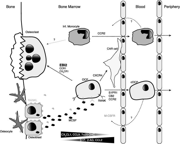
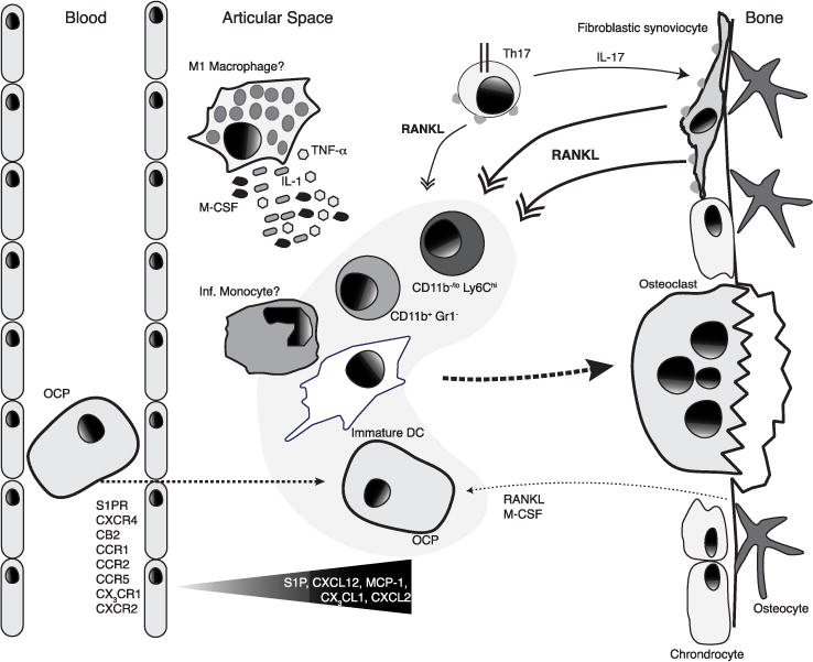
Rheumatoid arthritis (RA) is a chronic inflammatory autoimmune disease that primarily affects the joints. Self-reactive B and T lymphocytes cooperate to promote antibody responses against self proteins and are major drivers of disease. T lymphocytes also promote RA independently of B lymphocytes mainly through the production of key inflammatory cytokines, such as IL-17, that promote pathology. While the innate signals that initiate self-reactive adaptive immune responses are poorly understood, the disease is predominantly caused by inflammatory cellular infiltration and accumulation in articular tissues, and by bone erosions driven by bone-resorbing osteoclasts. Osteoclasts are giant multinucleated cells formed by the fusion of multiple myeloid cells that require short-range signals, such as the cytokines MCSF and RANKL, for undergoing differentiation. The recruitment and positioning of osteoclast precursors to sites of osteoclast differentiation by chemoattractants is an important point of control for osteoclastogenesis and bone resorption. Recently, the GPCR EBI2 and its oxysterol ligand 7a, 25 dihydroxycholesterol, were identified as important regulators of osteoclast precursor positioning in proximity to bone surfaces and of osteoclast differentiation under homeostasis. In chronic inflammatory diseases like RA, osteoclast differentiation is also driven by inflammatory cytokines such as TNFa and IL-1, and can occur independently of RANKL. Finally, there is growing evidence that the chemotactic signals guiding osteoclast precursors to inflamed articular sites contribute to disease and are of great interest. Furthering our understanding of the complex osteoimmune cell interactions should provide new avenues of therapeutic intervention for RA.
Keywords: Cell migration; Osteoclast precursors; Osteoclasts; Rheumatoid arthritis.
Conflict of interest statement
Conflict of Interest: E. Nevius, A.C. Gomes, and J.P. Pereira declare that they have no conflict of interest.
Figures
References
-
- Roadmap Epigenomics C, Kundaje A, Meuleman W, Ernst J, Bilenky M, Yen A, Heravi-Moussavi A, Kheradpour P, Zhang Z, Wang J, Ziller MJ, Amin V, Whitaker JW, Schultz MD, Ward LD, Sarkar A, Quon G, Sandstrom RS, Eaton ML, Wu YC, Pfenning AR, Wang X, Claussnitzer M, Liu Y, Coarfa C, Harris RA, Shoresh N, Epstein CB, Gjoneska E, Leung D, Xie W, Hawkins RD, Lister R, Hong C, Gascard P, Mungall AJ, Moore R, Chuah E, Tam A, Canfield TK, Hansen RS, Kaul R, Sabo PJ, Bansal MS, Carles A, Dixon JR, Farh KH, Feizi S, Karlic R, Kim AR, Kulkarni A, Li D, Lowdon R, Elliott G, Mercer TR, Neph SJ, Onuchic V, Polak P, Rajagopal N, Ray P, Sallari RC, Siebenthall KT, Sinnott-Armstrong NA, Stevens M, Thurman RE, Wu J, Zhang B, Zhou X, Beaudet AE, Boyer LA, De Jager PL, Farnham PJ, Fisher SJ, Haussler D, Jones SJ, Li W, Marra MA, McManus MT, Sunyaev S, Thomson JA, Tlsty TD, Tsai LH, Wang W, Waterland RA, Zhang MQ, Chadwick LH, Bernstein BE, Costello JF, Ecker JR, Hirst M, Meissner A, Milosavljevic A, Ren B, Stamatoyannopoulos JA, Wang T, Kellis M. Integrative analysis of 111 reference human epigenomes. Nature. 2015;518(7539):317–330. doi: 10.1038/nature14248. - DOI - PMC - PubMed
-
- Farh KK, Marson A, Zhu J, Kleinewietfeld M, Housley WJ, Beik S, Shoresh N, Whitton H, Ryan RJ, Shishkin AA, Hatan M, Carrasco-Alfonso MJ, Mayer D, Luckey CJ, Patsopoulos NA, De Jager PL, Kuchroo VK, Epstein CB, Daly MJ, Hafler DA, Bernstein BE. Genetic and epigenetic fine mapping of causal autoimmune disease variants. Nature. 2015;518(7539):337–343. doi: 10.1038/nature13835. - DOI - PMC - PubMed
Publication types
MeSH terms
Substances
Grants and funding
LinkOut - more resources
Full Text Sources
Other Literature Sources
Medical
Research Materials
