Comparative Study
. 2015 Oct 28;90(2):790-804.
doi: 10.1128/JVI.02258-15. Print 2016 Jan 15.

Herpes Simplex Virus 1 Infection of Tree Shrews Differs from That of Mice in the Severity of Acute Infection and Viral Transcription in the Peripheral Nervous System

Affiliations
Comparative Study

Herpes Simplex Virus 1 Infection of Tree Shrews Differs from That of Mice in the Severity of Acute Infection and Viral Transcription in the Peripheral Nervous System

Lihong Li et al. J Virol. .

Abstract

Studies of herpes simplex virus (HSV) infections of humans are limited by the use of rodent models such as mice, rabbits, and guinea pigs. Tree shrews (Tupaia belangeri chinensis) are small mammals indigenous to southwest Asia. At behavioral, anatomical, genomic, and evolutionary levels, tree shrews are much closer to primates than rodents are, and tree shrews are susceptible to HSV infection. Thus, we have studied herpes simplex virus 1 (HSV-1) infection in the tree shrew trigeminal ganglion (TG) following ocular inoculation. In situ hybridization, PCR, and quantitative reverse transcription-PCR (qRT-PCR) analyses confirm that HSV-1 latently infects neurons of the TG. When explant cocultivation of trigeminal ganglia was performed, the virus was recovered after 5 days of cocultivation with high efficiency. Swabbing the corneas of latently infected tree shrews revealed that tree shrews shed virus spontaneously at low frequencies. However, tree shrews differ significantly from mice in the expression of key HSV-1 genes, including ICP0, ICP4, and latency-associated transcript (LAT). In acutely infected tree shrew TGs, no level of ICP4 was observed, suggesting the absence of infection or a very weak, acute infection compared to that of the mouse. Immunofluorescence staining with ICP4 monoclonal antibody, and immunohistochemistry detection by HSV-1 polyclonal antibodies, showed a lack of viral proteins in tree shrew TGs during both acute and latent phases of infection. Cultivation of supernatant from homogenized, acutely infected TGs with RS1 cells also exhibited an absence of infectious HSV-1 from tree shrew TGs. We conclude that the tree shrew has an undetectable, or a much weaker, acute infection in the TGs. Interestingly, compared to mice, tree shrew TGs express high levels of ICP0 transcript in addition to LAT during latency. However, the ICP0 transcript remained nuclear, and no ICP0 protein could be seen during the course of mouse and tree shrew TG infections. Taken together, these observations suggest that the tree shrew TG infection differs significantly from the existing rodent models.

Importance: Herpes simplex viruses (HSVs) establish lifelong infection in more than 80% of the human population, and their reactivation leads to oral and genital herpes. Currently, rodent models are the preferred models for latency studies. Rodents are distant from primates and may not fully represent human latency. The tree shrew is a small mammal, a prosimian primate, indigenous to southwest Asia. In an attempt to further develop the tree shrew as a useful model to study herpesvirus infection, we studied the establishment of latency and reactivation of HSV-1 in tree shrews following ocular inoculation. We found that the latent virus, which resides in the sensory neurons of the trigeminal ganglion, could be stress reactivated to produce infectious virus, following explant cocultivation and that spontaneous reactivation could be detected by cell culture of tears. Interestingly, the tree shrew model is quite different from the mouse model of HSV infection, in that the virus exhibited only a mild acute infection following inoculation with no detectable infectious virus from the sensory neurons. The mild infection may be more similar to human infection in that the sensory neurons continue to function after herpes reactivation and the affected skin tissue does not lose sensation. Our findings suggest that the tree shrew is a viable model to study HSV latency.


Figures

FIG 1
FIG 1
PCR for HSV-1 DNA in tree shrew trigeminal ganglia 4 weeks after eye infection. For PCR detection of the viral genome, TG total DNA was extracted as described in Materials and Methods, followed by PCR detection of HSV-1-specific sequences using primers (see table in panel F). (A) PCR results from primer pair 1. Lanes 1 and 2, TGs from mock-infected tree shrews; lanes 3 through 10, infected tree shrew (TS) TGs; lane 11, HSV-infected Vero cells at 48 h postinfection (hpi) in culture; lane 12, double-distilled (DD) water as a PCR template control. Primer pair 1 gives a PCR product of 185 bp. (B and C) PCR products from the same set of templates as panel A but using primer sets 2 and 3, respectively. The PCR product was sequenced, and a Basic Alignment Search Tool (BLAST) search was performed to verify that the sequence was HSV. (D) Mouse (BALB/c) control. Lanes 1 and 2, mock-infected mouse TGs subjected to PCR using primer pair 1; lanes 3 to 6, PCR on TGs from four different mice infected with HSV-1 17+ strain; lane 7, PCR of Vero cells infected with HSV-1 17+ at 48 hpi; lane 8, PCR using DD water as a control. (E) Similar to panel D, except that primer pair 3 was used. (F) Table of the three pairs of primers used in the experiments. The orientation of the primer is indicated at the end of the primer name as follows: F, forward; R, reverse.
FIG 2
FIG 2
Explant cocultivation of tree shrew TG on RS1 cells. At 4 weeks or 60 days postinfection (p.i.), the animals (animal 80, 92, 62, and 76) were euthanized, TGs were removed from the animals and incubated with monolayers of rabbit skin cells (RS1). For a control, we also homogenized part of each TG and collected the supernatant after spinning down the tissue debris and incubated the supernatant with a monolayer of RS1 cells. The monolayers were inspected daily for signs of CPE until reactivation occurred. (A) Mock-infected tree shrew TG showed no sign of CPE during the entire time of incubation. (B) HSV-1 17+-infected tree shrew TG displayed CPE around the tree shrew tissue. (C) Mock-infected tree shrew TG underwent explant cocultivation, and no CPE was observed. (D) TGs from infected tree shrew at 60 days p.i. were cultured with RS1 cells. CPE is seen near TG tissue debris. (E) Statistical analysis of reactivation from latently infected tree shrews. There was 78% total reactivation. dpi, days postinfection.
FIG 3
FIG 3
In situ hybridization detection of LAT signal in HSV-1-infected TGs. RNA probes corresponding to the LAT intron region (positions 120384 to 121418) of HSV-1 were labeled with DigU. Dissected tree shrew TGs were embedded in paraffin and cut into sections 5 μm thick on a microtome (Leica RM2245 microtome). The sections were deparaffinized in xylene and a gradient concentration of ethanol. RNA in situ hybridization was performed as described in Materials and Methods. Each panel shows representative staining from groups of between three to six animals. (A) Mock-infected tree shrew TG showed no LAT signal. (B) The LAT signal was first detected by day 7 p.i. (yellow arrow). (C) By day 28, the LAT in situ signal was readily detected. (D) By 58 days postinfection (dpi), LAT signal persisted and remained strong.
FIG 4
FIG 4
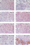
Immunohistochemistry using antibody against HSV-1 protein showed negative signals in tree shrew TGs during the acute phase of infection. (A) Mock-infected tree shrew (TS) TG subjected to the immunohistochemistry (IHC) procedure was negative for HSV-1 proteins. (B to D) HSV-1 17+-infected TS TGs display no HSV-1 antigen signals at 3, 5, and 7 days postinfection (dpi), respectively. (E and F) During the latent infection period, 4 and 8 weeks p.i. (27 and 53 days postinfection, respectively), tree shrew TG IHC was negative for HSV antigens. (G) Six BALB/c mice infected with the 17+ strain of HSV-1 showed no detected amount of HSV-1 antigen at 3 dpi. (H) Six BALB/c mice infected with HSV-1 17+ strain showed significant amounts of HSV-1 antigen at 5 dpi. The black arrows indicate large sensory neurons, the yellow arrows indicate smaller nonneuronal cells, and the red arrows show that the majority of HSV-1 antigen is located in the cytoplasm.
FIG 5
FIG 5
Immunohistochemistry detection of HSV-1 antigens in infected mouse and tree shrew CNS. The corneas of mice and tree shrews were inoculated with the McKrae strain of HSV-1 without ocular scarification. CNS tissues were dissected, sectioned, and processed for immunohistochemistry with anti-HSV-1 polyclonal antibodies. (A) Mock-infected mouse control. (B) Mouse CNS. The arrows point to clusters of infected neurons positive for HSV-1 antigens. (C) Tree shrew CNS mock-infected control. (D) HSV-1-infected tree shrew CNS. The arrows indicate neurons positive for HSV-1 antigens.
FIG 6
FIG 6
qRT-PCR detection of HSV-1 transcripts in infected animal TGs shows different expression profiles in mice and tree shrews. Mice and tree shrews infected with 17+ or McKrae strain of HSV-1 were euthanized and TGs were removed from the animals. RNAs from these samples were extracted for qRT-PCR analyses. (A) Six BALB/c mice were infected with the 17+ strain of HSV-1, and TGs were isolated at various days postinfection. qRT-PCR results for ICP0, ICP4, LAT, and UL36 were plotted. (B) Similar to panel A, except TGs from tree shrews infected with HSV-1 17+ strain were analyzed. (C) Similar to panel B except that the McKrae strain was used. (D) Primers used in the qRT-PCR detection of viral transcripts.
FIG 7
FIG 7
Immunofluorescence staining of ICP4 in mouse and tree shrew TGs. BALB/c mice and tree shrews were infected with the 17+ strain of HSV-1, and at 3 dpi (mouse and tree shrew), 5 dpi (mouse and tree shrew), and 7 dpi (tree shrew), the animals were euthanized and dissected to obtain TGs, which were paraffin embedded and sectioned on a microtome before immunofluorescence staining with ICP4 antibody. (A) Mock-infected BALB/c mouse TG. No positive signal was detected. Arrows indicate neuronal nucleus. (B) HSV-1 17+-infected mouse at 3 days postinfection showed no ICP4 signal. (C) At 5 dpi, the ICP4 signal was readily detected in large neuronal cells in the TG. The arrows point to large neuronal nuclei, where ICP4 signal could be seen. (D) At 5 dpi, ICP4 stained not only the neuronal nucleus (white arrows) but also smaller nonneuronal cells (yellow arrows). (E) TG section in a mock-infected tree shrew. Arrows point to neuronal cell nuclei. (F) Infected TS TG at 3 dpi. No ICP4 signal was seen. (G) Infected TS TG at 5 dpi. No ICP4 signal was detected. (H) HSV-1 17+-infected tree shrew TG section at 7 dpi. No ICP4 signal was detected.
FIG 8
FIG 8
In situ hybridization detection of ICP0 signals in mouse and tree shrew TGs. (A) During the acute stage of infection (5 dpi using HSV-1 17+), LAT transcript is readily detected using RNA probe against the first exon of LAT. (B) Compared to mouse TG, LAT exon is undetected at the same time point in infected tree shrew TGs. (C) ICP0 is expressed in HSV-1 17+-infected mouse TG at 5 dpi. (D) The ICP0 transcript is undetected in most HSV-1 17+-infected mouse TGs during latency. (E) In acutely HSV-1 17+-infected tree shrew TG, no ICP0 was detected at 5 dpi. (F) In latently HSV-1 17+-infected tree shrew TG, ICP0 could be detected at day 58 postinfection. (G) Unlike strain 17+, the McKrae strain-infected tree shrew TGs show ICP0 expression at 5 dpi. (H) In latent TGs infected with McKrae strain, ICP0 is readily detected. (I) In situ hybridization of ICP0 in HSV-1-infected BJ cells. (J) In situ hybridization of LAT exon in HSV-1-infected BJ cells.
FIG 9
FIG 9
HSV-1 virus is not detected from tree shrew TGs during the acute stage of infection. TGs were dissected from mock-infected mice and tree shrews and infected mice and tree shrews. The TGs from infected animals were dissected 3, 5, or 7 days after inoculating the eyes of the animal with the 17+ strain of HSV-1, and the TGs were homogenized. The supernatant was incubated with RS1 cells to observe CPE and also incubated with RS cells on 96-well plates to calculate viral titer. (A) Mock-infected mouse TG control. A CPE was detected after 3 days. Day 5 showed stronger CPE than day 7 postinfection. (B) No CPE was detected with tree shrew TG supernatants. (C) Titers of HSV-1 loads were measured for infected mouse and tree shrew TGs. Each point represents the geometric mean titer determined from four individual ganglia at the indicated time (days) postinfection. The titers are plotted on a logarithmic scale as the number of PFU per trigeminal ganglia (PFU/TG). (D) Relative viral genome copy number in mouse and tree shrew TGs were detected by real-time PCR. Mice or tree shrews were infected as described above. Viral DNA levels at each time point were quantified relative to the 13 dpi sample by the ΔΔCT method as follows: ΔΔCT = (CTtestCTreference) − (CT13 dpiCT13 dpi β-actin) where test, reference, 13 dpi, and 13 dpi β-actin refer to the samples (e.g., CTtest is the CT of the test sample). The fold enrichment value is 2−ΔΔCT. The values are means ± standard errors (SE) (error bar) of ≥3 samples per data point.

References

    1. Steiner I, Benninger F. 2013. Update on herpes virus infections of the nervous system. Curr Neurol Neurosci Rep 13:414. doi:10.1007/s11910-013-0414-8. - DOI - PubMed
    1. Webre JM, Hill JM, Nolan NM, Clement C, McFerrin HE, Bhattacharjee PS, Hsia V, Neumann DM, Foster TP, Lukiw WJ, Thompson HW. 2012. Rabbit and mouse models of HSV-1 latency, reactivation, and recurrent eye diseases. J Biomed Biotechnol 2012:612316. doi:10.1155/2012/612316. - DOI - PMC - PubMed
    1. Stuart PM, Keadle TL. 2012. Recurrent herpetic stromal keratitis in mice: a model for studying human HSK. Clin Dev Immunol 2012:728480. doi:10.1155/2012/728480. - DOI - PMC - PubMed
    1. Dasgupta G, BenMohamed L. 2011. Of mice and not humans: how reliable are animal models for evaluation of herpes CD8+-T cell-epitopes-based immunotherapeutic vaccine candidates? Vaccine 29:5824–5836. doi:10.1016/j.vaccine.2011.06.083. - DOI - PMC - PubMed
    1. Bernstein DI, Stanberry LR, Harrison CJ, Kappes JC, Myers MG. 1986. Antibody response, recurrence patterns and subsequent herpes simplex virus type 2 (HSV-2) reinfection following initial HSV-2 infection of guinea pigs: effects of acyclovir. J Gen Virol 67:1601–1612. doi:10.1099/0022-1317-67-8-1601. - DOI - PubMed

Publication types