Intramuscular schwannoma: clinical and magnetic resonance imaging features
- PMID: 26512147
- PMCID: PMC4613930
- DOI: 10.11622/smedj.2015151
Intramuscular schwannoma: clinical and magnetic resonance imaging features
Abstract
Introduction: Schwannomas that arise within the muscle plane are called intramuscular schwannomas. The low incidence of these tumours and the lack of specific clinical features make preoperative diagnosis difficult. Herein, we report our experience with intramuscular schwannomas. We present details of the clinical presentation, radiological diagnosis and management of these tumours.
Methods: Between January 2011 and December 2013, 29 patients were diagnosed and treated for histologically proven schwannoma at the National University Hospital, Singapore. Among these 29 patients, eight (five male, three female) had intramuscular schwannomas.
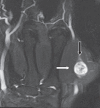
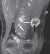
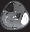
Results: The mean age of the eight patients was 40 (range 27-57) years. The most common presenting feature was a palpable mass. The mean interval between surgical treatment and the onset of clinical symptoms was 17.1 (range 4-72) months. Six of the eight tumours (75.0%) were located in the lower limb, while 2 (25.0%) were located in the upper limb. None of the patients had any preoperative neurological deficits. Tinel's sign was present in one patient. Magnetic resonance (MR) imaging showed that the findings of split-fat sign, low signal margin and fascicular sign were present in all patients. The entry and exit sign was observed in 4 (50.0%) patients, a hyperintense rim was observed in 7 (87.5%) patients and the target sign was observed in 5 (62.5%) patients. All patients underwent microsurgical excision of the tumour and none developed any postoperative neurological deficits.
Conclusion: Intramuscular schwannomas demonstrate the findings of split-fat sign, low signal margin and fascicular sign on MR imaging. These findings are useful for the radiological diagnosis of intramuscular schwannoma.
Keywords: intramuscular; magnetic resonance imaging; schwannoma.
Figures


References
-
- Kransdorf MJ. Benign soft-tissue tumors in a large referral population: distribution of specific diagnoses by age, sex, and location. AJR Am J Roentgenol. 1995;164:395–402. - PubMed
-
- Enzinger FM, Weiss SW, editors. Soft tissue tumors. St Louis, MO: Mosby; 1995. Benign tumors of peripheral nerves; pp. 821–88.
-
- Murphey MD, Smith WS, Smith SE, Kransdorf MJ, Temple HT. From the archives of the AFIP. Imaging of musculoskeletal neurogenic tumors: radiologic-pathologic correlation. Radiographics. 1999;19:1253–80. - PubMed
-
- Shimose S, Sugita T, Kubo T, et al. Major-nerve schwannomas versus intramuscular schwannomas. Acta Radiol. 2007;48:672–7. - PubMed
-
- Kwon BC, Baek GH, Chung MS, et al. Intramuscular neurilemoma. J Bone Joint Surg Br. 2003;85:723–5. - PubMed
MeSH terms
LinkOut - more resources
Full Text Sources
Other Literature Sources
Medical

