

Moving back: The radiation dose received from lumbar spine quantitative fluoroscopy compared to lumbar spine radiographs with suggestions for dose reduction
- PMID: 26512196
- PMCID: PMC4579040
- DOI: 10.1016/j.radi.2014.03.010
Moving back: The radiation dose received from lumbar spine quantitative fluoroscopy compared to lumbar spine radiographs with suggestions for dose reduction
Abstract
Purpose: Quantitative fluoroscopy is an emerging technology for assessing continuous inter-vertebral motion in the lumbar spine, but information on radiation dose is not yet available. The purposes of this study were to compare the radiation dose from quantitative fluoroscopy of the lumbar spine with lumbar spine radiographs, and identify opportunities for dose reduction in quantitative fluoroscopy.
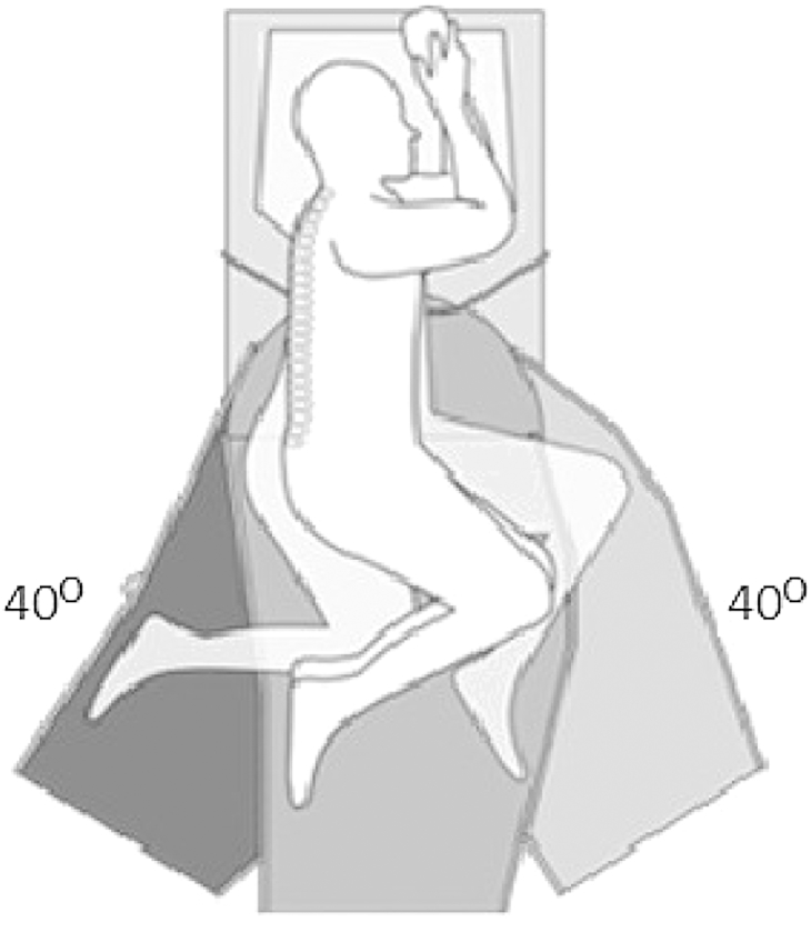
Methods: Internationally reported dose area product (DAP) and effective dose data for lumbar spine radiographs were compared with the same for quantitative fluoroscopy and with data from a local hospital for functional radiographs (weight bearing AP, lateral, and/or flexion and extension) (n = 27). The effects of procedure time, age, weight, height and body mass index on the fluoroscopy dose were determined by multiple linear regression using SPSS v19 software (IBM Corp., Armonck, NY, USA).
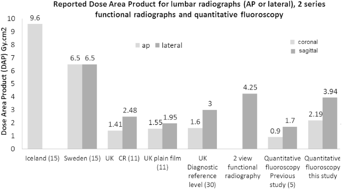
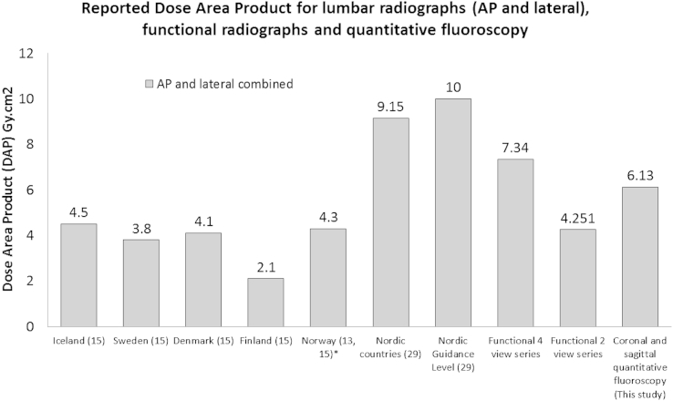
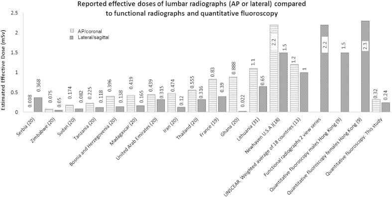
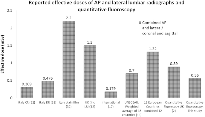
Results and conclusion: The effective dose (and therefore the estimated risk) for quantitative fluoroscopy is 0.561 mSv which is lower than in most published data for lumbar spine radiography. The dose area product (DAP) for sagittal (flexion + extension) quantitative fluoroscopy is 3.94 Gy cm2 which is lower than local data for two view (flexion and extension) functional radiographs (4.25 Gy cm2), and combined coronal and sagittal dose from quantitative fluoroscopy (6.13 Gy cm2) is lower than for four view functional radiography (7.34 Gy cm2). Conversely DAP for coronal and sagittal quantitative fluoroscopy combined (6.13 Gy cm2) is higher than that published for both lumbar AP or lateral radiographs, with the exception of Nordic countries combined data. Weight, procedure time and age were independently positively associated with total dose, and height (after adjusting for weight) was negatively associated, thus as height increased, the DAP decreased.
Keywords: Continuous motion; Flexion-extension; Inter-vertebral; Low back pain; Movement disorders; Spine kinematics.
Figures
References
-
- Mellor F.E., Muggleton J.M., Bagust J., Mason W.M.A., Thomas P.W., Breen A.C. Midlumbar lateral flexion stability measured in healthy volunteers by in-vivo fluoroscopy. Spine. 2009;34(22):E811–E817. - PubMed
-
- Leone A., Cassar-Pullicino V., Gugliemli G., Bonomo L. Degenerative lumbar intervertebral instability: what is it and how does imaging contribute? Skelet Radiol. 2009;38(6):529–533. - PubMed
-
- Mayer R.S., Chen I.H., Lavender S.A., Trafimow J.H., Andersson G.B. Variance in the measurement of sagittal lumbar spine range of motion among examiners, subjects, and instruments. Spine. 1995;20(13):1489–1493. - PubMed
LinkOut - more resources
Full Text Sources
Other Literature Sources
