Review
. 2015 Oct 23;16(10):25392-432.
doi: 10.3390/ijms161025392.

Changes in Regenerative Capacity through Lifespan

Affiliations
Review

Changes in Regenerative Capacity through Lifespan

Maximina H Yun. Int J Mol Sci. .

Abstract

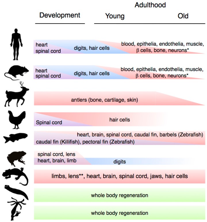
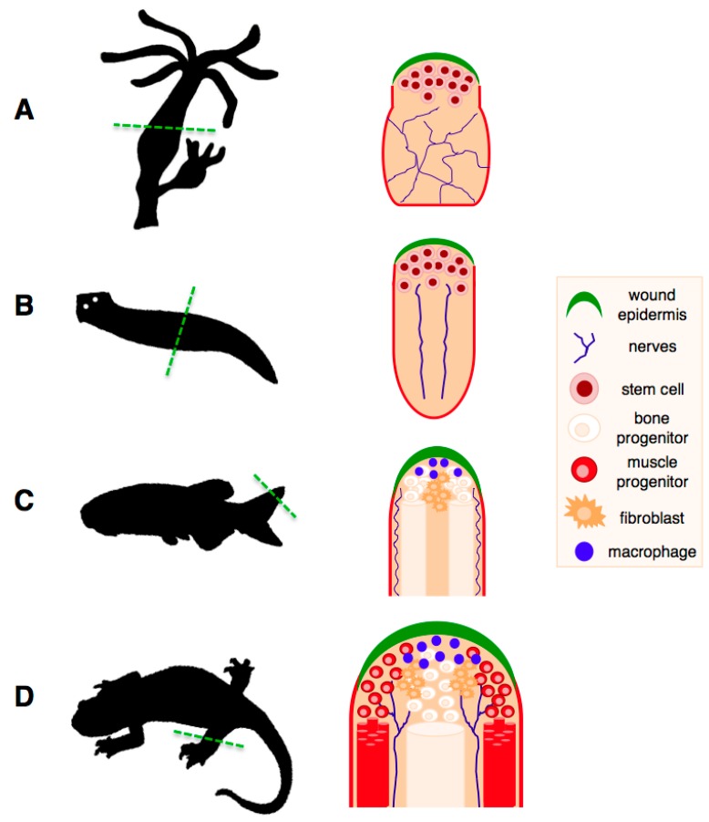
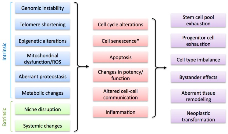
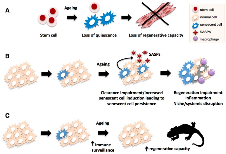
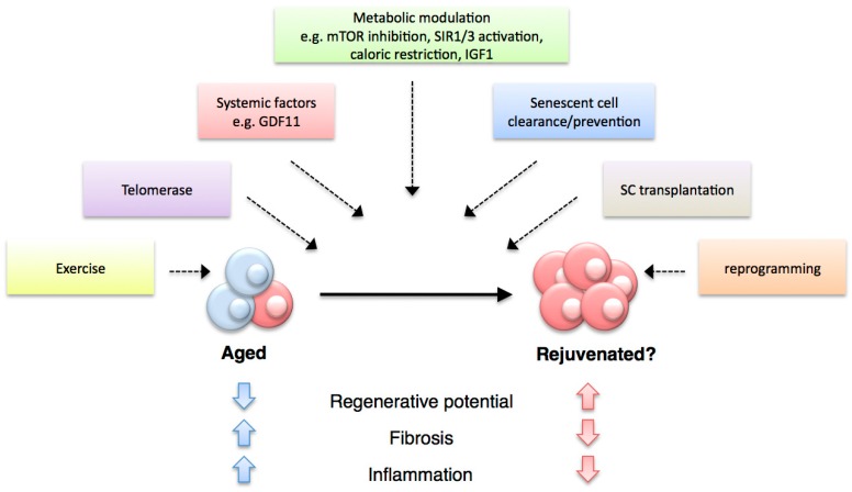
Most organisms experience changes in regenerative abilities through their lifespan. During aging, numerous tissues exhibit a progressive decline in homeostasis and regeneration that results in tissue degeneration, malfunction and pathology. The mechanisms responsible for this decay are both cell intrinsic, such as cellular senescence, as well as cell-extrinsic, such as changes in the regenerative environment. Understanding how these mechanisms impact on regenerative processes is essential to devise therapeutic approaches to improve tissue regeneration and extend healthspan. This review offers an overview of how regenerative abilities change through lifespan in various organisms, the factors that underlie such changes and the avenues for therapeutic intervention. It focuses on established models of mammalian regeneration as well as on models in which regenerative abilities do not decline with age, as these can deliver valuable insights for our understanding of the interplay between regeneration and aging.

Keywords: aging; axolotl; newt; planaria; regeneration; reprogramming; senescence; stem cells; zebrafish.


Figures

Figure 1
Figure 1
Variation in regenerative capacity through phylogeny, ontogeny and aging. * Note that the ability to regenerate the indicated systems is present in most other animal groups; ** Lens regeneration is observed throughout lifespan in newts, but it can only occur during a limited developmental window in axolotls.
Figure 2
Figure 2
Regeneration of complex structures in classic regeneration models. (A) Regeneration of a hydra polyp following amputation across the body stalk. Regeneration takes place through mobilisation and activation of multipotent endodermal and ectodermal stem cell populations; (B) Regeneration of a planarian flatworm following bisection. This process takes place through recruitment of pluripotent stem cells, termed “neoblasts”, which are present throughout the animal and carry out tissue maintenance functions. A single clonogenic neoblast is capable of regenerating an entire organism; (C) Regeneration of the zebrafish fin. Upon amputation of the fin, differentiated cells at the amputation plane undergo dedifferentiation and proliferate to form a pool of progenitors called a blastema, which will then undergo growth and redifferentiation into the new fin tissues; (D) Salamander limb regeneration depends, as in the zebrafish case, on the dedifferentiation of mature cells from the tissues at the amputation plane. Unlike the zebrafish fin, which grows continuously, salamander regeneration takes place in the context of mature adult tissues. Both in zebrafish and salamanders, the dedifferentiation process generates progenitors of limited potential, which can only regenerate their tissues of origin. The wound epithelium, nerve supply and macrophages are critical components of the regenerating niche, without which regeneration cannot proceed. Adapted from Brockes et al. [48].
Figure 3
Figure 3
Factors that alter regenerative capacity upon aging. Multiple cell-intrinsic and cell-extrinsic factors are associated with the decline of regenerative capacity during aging (left). These impact on regenerative cell types by altering key cellular processes (middle), leading to various negative outcomes that result in regeneration impairment (right). * Cell senescence can be considered an intrinsic and, potentially, extrinsic factor (see Section 4).
Figure 4
Figure 4
Impact of cellular senescence on regenerative processes during aging. Senescent cells accumulate in most organisms as they age, and this can negatively affect various regenerative processes. In the case of aged mammalian stem cells such as satellite cells (A), an age-associated switch to cellular senescence leads to loss of quiescence resulting in impaired regenerative ability; In addition to direct effects on regenerative capacity, senescent cells could promote tissue disruption, inflammation and niche or systemic alterations (B) through their phenotype (SASP), leading to further regenerative impairment; Senescent cell accumulation through aging can result from increases in stimuli that trigger cellular senescence (e.g., ROS, DNA damage, telomere attrition) or impairments in the immune-mediated clearance mechanism. In contrast to mammals, salamanders (C) have highly efficient mechanisms of senescence immunesurveillance which prevent senescent cell accumulation and could support their extensive regenerative abilities through lifespan.
Figure 5
Figure 5
Therapeutic strategies for improving regenerative capacity.

References

    1. Sharpless N.E., DePinho R.A. Telomeres, stem cells, senescence, and cancer. J. Clin. Investig. 2004;113:160–168. doi: 10.1172/JCI20761. - DOI - PMC - PubMed
    1. Finch C.E. Longevity, Senescence and the Genome. The University of Chicago Press; Chicago, IL, USA: 1994.
    1. Lopez-Otin C., Blasco M.A., Partridge L., Serrano M., Kroemer G. The hallmarks of aging. Cell. 2013;153:1194–1217. doi: 10.1016/j.cell.2013.05.039. - DOI - PMC - PubMed
    1. Seifert A.W., Voss S.R. Revisiting the relationship between regenerative ability and aging. BMC Biol. 2013;11:2. doi: 10.1186/1741-7007-11-2. - DOI - PMC - PubMed
    1. Porrello E.R., Mahmoud A.I., Simpson E., Hill J.A., Richardson J.A., Olson E.N., Sadek H.A. Transient regenerative potential of the neonatal mouse heart. Science. 2011;331:1078–1080. doi: 10.1126/science.1200708. - DOI - PMC - PubMed

Publication types