Review
. 2016 Mar 3;12(3):806-28.
doi: 10.1080/21645515.2015.1102804.

Peptide/protein vaccine delivery system based on PLGA particles

Affiliations
Review

Peptide/protein vaccine delivery system based on PLGA particles

Mojgan Allahyari et al. Hum Vaccin Immunother. .

Abstract

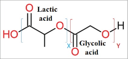
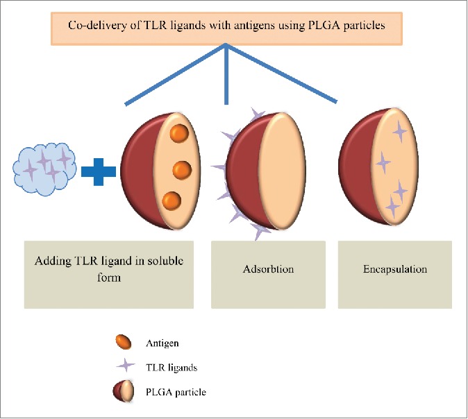
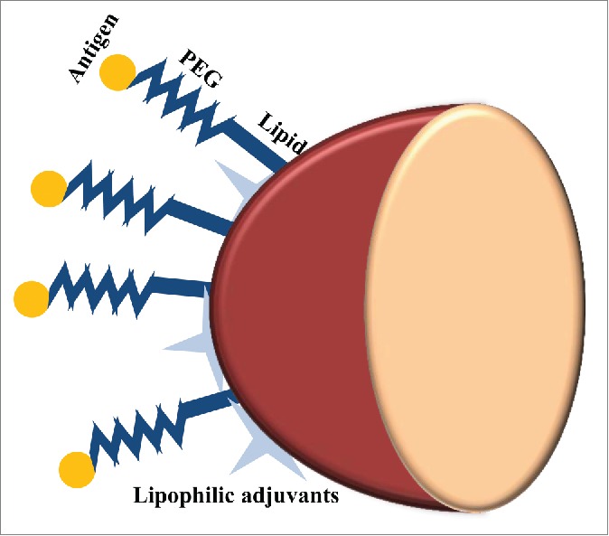
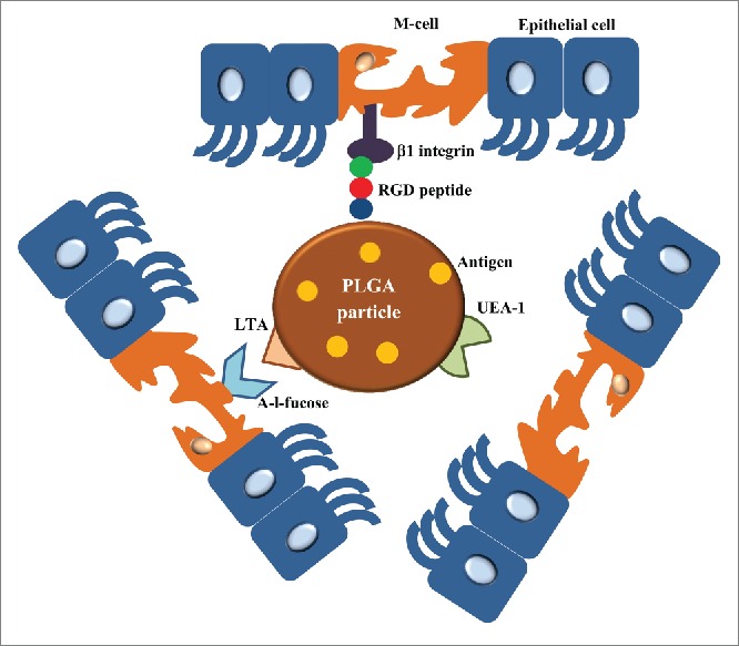
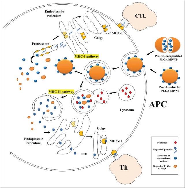
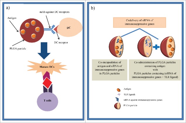
Due to the excellent safety profile of poly (D,L-lactide-co-glycolide) (PLGA) particles in human, and their biodegradability, many studies have focused on the application of PLGA particles as a controlled-release vaccine delivery system. Antigenic proteins/peptides can be encapsulated into or adsorbed to the surface of PLGA particles. The gradual release of loaded antigens from PLGA particles is necessary for the induction of efficient immunity. Various factors can influence protein release rates from PLGA particles, which can be defined intrinsic features of the polymer, particle characteristics as well as protein and environmental related factors. The use of PLGA particles encapsulating antigens of different diseases such as hepatitis B, tuberculosis, chlamydia, malaria, leishmania, toxoplasma and allergy antigens will be described herein. The co-delivery of antigens and immunostimulants (IS) with PLGA particles can prevent the systemic adverse effects of immunopotentiators and activate both dendritic cells (DCs) and natural killer (NKs) cells, consequently enhancing the therapeutic efficacy of antigen-loaded PLGA particles. We will review co-delivery of different TLR ligands with antigens in various models, highlighting the specific strengths and weaknesses of the system. Strategies to enhance the immunotherapeutic effect of DC-based vaccine using PLGA particles can be designed to target DCs by functionalized PLGA particle encapsulating siRNAs of suppressive gene, and disease specific antigens. Finally, specific examples of cellular targeting where decorating the surface of PLGA particles target orally administrated vaccine to M-cells will be highlighted.

Keywords: PLGA particle; TLR ligands; delivery system; micro/nano particles; protein/peptide vaccine.


Figures

Figure 1.
Figure 1.
PLGA polymer structure, PLGA a copolymer of poly lactic acid (PLA) and poly glycolic acid (PGA). X and Y indicate the number of each unit repeats.
Figure 2.
Figure 2.
Flow diagrams of single (a) and double (b) emulsion solvent evaporation methods. a) Single emulsion solvent evaporation (Oil/Water emulsion) flow diagram. Oil phase (O phase), Oil in water phase (O/W phase), microsphere (MS), b) Double emulsion solvent evaporation (Water/Oil/Water emulsion) flow diagram. Water in oil phase (W/O phase), water in oil in water (W/O/W), microsphere (MS).
Figure 3.
Figure 3.
Schematic illustration of the single emulsion evaporation method.
Figure 4.
Figure 4.
Flow diagram of PLGA polymer related factor affecting on PLGA particle degradation.
Figure 5.
Figure 5.
Co-delivery of immunepotentiators and antigens in PLGA particles. To enhance the therapeutic efficacy of PLGA particles, different TLR ligands (e.g., CpG, poly(I:C) and MPLA) can be adsorbed, encapsulated or added into soluble form along with antigen-loaded PLGA particles.
Figure 6.
Figure 6.
Pathogen-mimicking PLGA particles as vaccine delivery platform. In order to avoid antigen denaturation that can be caused by encapsulation process and preserve the structure and surface chemistry of antigens, they are conjugated to the PEGylated lipid shell of PLGA particles. Lipophilic molecular danger signals such as MPLA and αGC can also be incorporated into the surface of these lipid-enveloped PLGA particles.
Figure 7.
Figure 7.
Different ways to target PLGA particles to M-cells. The surface of PLGA particles can be grafted with RGD peptide or LTA which are interacted with β1 integrin and α-l-fucose presented in the surface of M-cells, respectively. UEA-1 is another M-cell targeting agent for oral-mucosal vaccine delivery. Using these M cell targeting agent-anchored PLGA particles, the orally administrated antigens loaded in PLGA particles are targeted to M-cells to generate mucosal immunity.
Figure 8.
Figure 8.
Schematic presentation of intracellular trafficking of protein/peptide - loaded PLGA particles in antigen presenting cells (mostly dendritic cell). Desired protein can be adsorbed on or encapsulated into PLGA particles, hence due to simplification only protein- adsorbed PLGA particles is shown. PLGA particles are taken up by endocytosis following binding to DC membrane. Antigens delivered by PLGA particles undergo 2 distinct intracellular pathways. In phagosome to cytosol pathway of cross-presentation, PLGA particles escape from the endosome, degrade in cytoplasm, and loaded protein is release gradually, and continue the MHC class I pathway. Secondly, degradation of protein/peptide-loaded PLGA particle happens inside endosome due to its acidic pH, thus the antigenic peptides derived from degradation can be presented via MHC class II pathway. Antigen presenting cells: APC; TCL: T cytotoxic lymphocyte; Th: T-helper lymphocyte.
Figure 9.
Figure 9.
Schematic illustration of strategies to enhance the therapeutic efficacy of DC-based vaccines via PLGA particles. (a) PLGA particles are linked to specific antibodies against DC receptors (e.g., DEC-205 and CD11c) to target vaccine components which are loaded in these particles specifically to DCs. Therefore, DC maturation and consequently CD8+ activation are occurred. (b) In another strategy, siRNA of suppressive gene can either co-encapsulate in PLGA particles containing antigen or encapsulate in distinct PLGA particle with TLR ligands and co-administer with antigen loaded-PLGA particles. Then, DCs are activated by these PLGA particles and antigen specific immunity is induced.

References

    1. Jain S, O'Hagan DT, Singh M. The long-term potential of biodegradable poly(lactide-co-glycolide) microparticles as the next-generation vaccine adjuvant. Expert Rev Vaccines 2011; 10:1731-42; PMID:22085176; http://dx.doi.org/10.1586/erv.11.126 - DOI - PubMed
    1. Amjadi I, Rabiee M, Hosseini MS. Anticancer Activity of Nanoparticles Based on PLGA and its Co-polymer: In-vitro Evaluation. Iran J Pharm Res 2013; 12:623-34; PMID:24523742 - PMC - PubMed
    1. Makadia HK, Siegel SJ. Poly lactic-co-glycolic acid (PLGA) as biodegradable controlled drug delivery carrier. Polymers 2011; 3:1377-97; PMID:22577513; http://dx.doi.org/10.3390/polym3031377 - DOI - PMC - PubMed
    1. Semete B, Booysen L, Lemmer Y, Kalombo L, Katata L, Verschoor J, Swai HS. In vivo evaluation of the biodistribution and safety of PLGA nanoparticles as drug delivery systems. Nanomedicine 2010; 6:662-71; http://dx.doi.org/10.1016/j.nano.2010.02.002 - DOI - PubMed
    1. Mahboubian A, Hashemein SK, Moghadam S, Atyabi F, Dinarvand R. Preparation and in-vitro evaluation of controlled release PLGA microparticles containing triptoreline. Iran J Pharm Res 2010; 9:369; PMID:24381601 - PMC - PubMed

MeSH terms