. 2015 Nov 17;6(36):39342-56.
doi: 10.18632/oncotarget.5745.

Celecoxib increases lung cancer cell lysis by lymphokine-activated killer cells via upregulation of ICAM-1

Affiliations

Celecoxib increases lung cancer cell lysis by lymphokine-activated killer cells via upregulation of ICAM-1

Melina Schellhorn et al. Oncotarget. .

Abstract

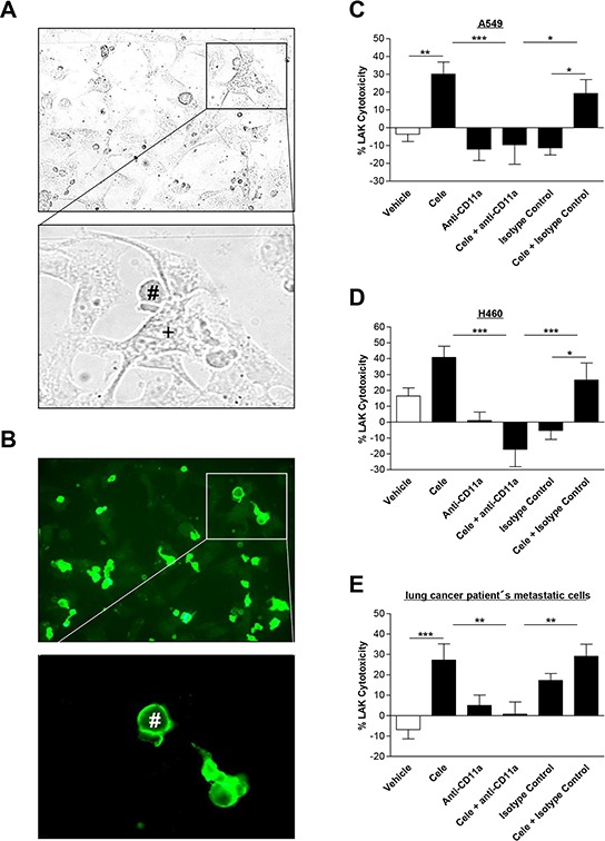
The antitumorigenic mechanism of the selective cyclooxygenase-2 (COX-2) inhibitor celecoxib is still a matter of debate. Using lung cancer cell lines (A549, H460) and metastatic cells derived from a lung cancer patient, the present study investigates the impact of celecoxib on the expression of intercellular adhesion molecule 1 (ICAM-1) and cancer cell lysis by lymphokine-activated killer (LAK) cells. Celecoxib, but not other structurally related selective COX-2 inhibitors (i.e., etoricoxib, rofecoxib, valdecoxib), was found to cause a substantial upregulation of ICAM-1 protein levels. Likewise, ICAM-1 mRNA expression was increased by celecoxib. Celecoxib enhanced the susceptibility of cancer cells to be lysed by LAK cells with the respective effect being reversed by a neutralizing ICAM-1 antibody. In addition, enhanced killing of celecoxib-treated cancer cells was reversed by preincubation of LAK cells with an antibody to lymphocyte function associated antigen 1 (LFA-1), suggesting intercellular ICAM-1/LFA-1 crosslink as crucial event within this process. Finally, celecoxib elicited no significant increase of LAK cell-mediated lysis of non-tumor bronchial epithelial cells, BEAS-2B, associated with a far less ICAM-1 induction as compared to cancer cells. Altogether, our data demonstrate celecoxib-induced upregulation of ICAM-1 on lung cancer cells to be responsible for intercellular ICAM-1/LFA-1 crosslink that confers increased cancer cell lysis by LAK cells. These findings provide proof for a novel antitumorigenic mechanism of celecoxib.

Keywords: celecoxib; immune surveillance; intercellular adhesion molecule 1; lung cancer cells; lymphokine-activated killer cells.


Conflict of interest statement

CONFLICTS OF INTEREST

The authors indicate no potential conflicts of interest.

Figures

Figure 1
Figure 1. Effect of celecoxib and other selective COX-2 inhibitors on ICAM-1 protein expression in human lung cancer cells
Left panels: Concentration-dependent impact of celecoxib on ICAM-1 protein expression in A549 A. H460 B. and lung cancer patient's metastatic cells C. Cells were incubated with celecoxib at the indicated concentrations for 48 h. Histograms above selected blots represent means ± SEM obtained from densitometric analysis of n = 4 (A, C) or n = 3 (B) blots. Right panels: Influence of selective COX-2 inhibitors on ICAM-1 protein expression in A549 D. H460 E. and lung cancer patient's metastatic cells F. Tumor cells were treated with 30 μM celecoxib (Cele), etoricoxib (Etori), rofecoxib (Rofe), valdecoxib (Valde) or vehicle for 48 h. Histograms above selected blots represent means ± SEM obtained from densitometric analysis of n = 4 (D, E, F) blots. *P < 0.05, **P < 0.01, ***P < 0.001; one-way ANOVA plus post hoc Dunnett test.
Figure 2
Figure 2. Time-dependent impact of celecoxib on ICAM-1 expression in A549, H460 and lung cancer patient's metastatic cells
A–C. Western blot analysis of celecoxib's (30 μM) effect on ICAM-1 protein expression over a 48-h incubation period. Values are means ± SEM obtained from densitometric analysis of n = 3 blots. *P < 0.05, **P < 0.01, ***P < 0.001 vs. corresponding vehicle control of the respective ICAM-1 analysis; Student's t test. D–F. Real-time RT-PCR analysis of the impact of 30 μM celecoxib on ICAM-1 mRNA expression over a 48-h incubation period. Values are means ± SEM of n = 4 experiments. *P < 0.05, **P < 0.01, ***P < 0.001 vs. corresponding vehicle control of the respective ICAM-1 analysis; Student's t test.
Figure 3
Figure 3. Effect of celecoxib on LAK cell-mediated tumor cell killing
A series of scanning electron micrographs (A. left panels) visualizes the interactions between LAK cells and tumor cells. Electron micrographs at lower magnification show that LAK cells firmly attach to the spread A549 tumor cells with their processes (upper two panels). In addition, immunolabelling with LFA-1 antibody and a secondary antibody coupled to 15 nm colloidal gold was used to mark LAK cells. The gold labelling is visible as bright dots in the electron micrographs at higher magnifications (lower two panels with inserts) and decorates the cell body (second to last panel) as well as the processes of the LAK cell (lowermost panel, corresponding to the boxed area in the low magnification second from above panel). Note that the 15 nm gold labeling is confined to the processes of the LAK cell (open arrows) but is absent from the intermingled filopodia of the underlying tumor cell (filled arrows). Right panels: Concentration-dependent impact of celecoxib on LAK cell-mediated killing of A549 B. H460 C. or lung cancer patient's metastatic cells D. Tumor cells were incubated with celecoxib at the indicated concentrations for 48 h. Subsequently, these cells were co-incubated with LAK cells for 6 h. Values are means ± SEM of n = 24 (B, 6 donors), n = 28 (C, 7 donors) or n = 20 (D, 5 donors) experiments. *P < 0.05, ***P < 0.001 vs. corresponding vehicle control; one-way ANOVA plus post hoc Dunnett test.
Figure 4
Figure 4. Impact of celecoxib on cytotoxic activity of LAK cells
The respective subgroups indicate lysis of cancer cells when LAK cells were preincubated for 48 h in medium (white bars), medium containing vehicle (grey bars) or 30 μM celecoxib (black bars) prior to co-culturing with cancer cells. As LAK cell medium RPMI 1640 supplemented with 10% heat-inactivated FCS, 100 U/ml penicillin, 100 μg/ml streptomycin and 10 ng/ml IL-2 was used. Cytotoxicity was analyzed following a subsequent 6-h co-incubation of LAK cells with cancer cells. Values are means ± SEM of n = 12 (A. 3 donors), n = 20 (B. 5 donors) or n = 16 (C. 4 donors) experiments. **P < 0.01, ***P < 0.001 vs. corresponding vehicle control; Student's t test.
Figure 5
Figure 5. Effect of a neutralizing ICAM-1 antibody on cytotoxic lysis of cancer cells by LAK cells
A549 A. H460 B. and lung cancer patient's metastatic cells C. were incubated with vehicle or 30 μM celecoxib for 48 h. Before starting cytotoxicity assay cancer cells were pre-incubated with an ICAM-1 antibody (1 μg/ml) for 2 h. An isotype control antibody (1 μg/ml) was used as negative control. Values are means ± SEM of n = 24 (A, 6 donors), n = 16 (B, 4 donors) or n = 20 (C, 5 donors) experiments. *P < 0.05, **P < 0.01, ***P < 0.001; one-way ANOVA plus post hoc Bonferroni test.
Figure 6
Figure 6. Effect of a CD11a (LFA-1) neutralizing antibody on interaction of cancer cells with LAK cells
Light microscopy picture A. presents interaction between tumor (+) and LAK cells (#), whereas fluorescence microscopy pictures show immunocytochemical staining of LFA-1 expression on the surface of the immune cells (B. green fluorescence). Right panels: Influence of CD11a (LFA-1) antibody on cytotoxic lysis of cancer cells by LAK cells. A549 C. H460 D. and lung cancer patient's metastatic cells E. were incubated with vehicle or 30 μM celecoxib for 48 h. LAK cells were pre-incubated with a CD11a antibody (1 μg/ml) or an isotype control antibody (1 μg/ml) as negative control for 2 h before cytotoxicity assay was started. Values are means ± SEM of n = 20 (C, D, 5 donors) or n = 28 (E, 7 donors) experiments. *P < 0.05, **P < 0.01, ***P < 0.001; one-way ANOVA plus post hoc Bonferroni test.
Figure 7
Figure 7. Concentration-dependent effect of celecoxib on ICAM-1 protein expression in human bronchial epithelial cells (BEAS-2B) and on cytotoxic lysis of BEAS-2B cells by LAK cells
BEAS-2B cells were incubated with celecoxib at the indicated concentrations for 48 h. Histogram A. above the selected blot represents means ± SEM obtained from densitometric analysis of n = 5 blots. Values in B. are means ± SEM of n = 20 (5 donors) experiments. One-way ANOVA plus post hoc Dunnett test revealed no significant effect of celecoxib vs. vehicle.

References

    1. McAdam BF, Catella-Lawson F, Mardini IA, Kapoor S, Lawson JA, FitzGerald GA. Systemic biosynthesis of prostacyclin by cyclooxygenase (COX)-2: the human pharmacology of a selective inhibitor of COX-2. Proc Natl Acad Sci USA. 1999;96:272–277. - PMC - PubMed
    1. Steinbach G, Lynch PM, Phillips RK, Wallace MH, Hawk E, Gordon GB, Wakabayashi N, Saunders B, Shen Y, Fujimura T, Su LK, Levin B, Godio L, et al. The effect of celecoxib, a cyclooxygenase-2 inhibitor, in familial adenomatous polyposis. N Engl J Med. 2000;342:1946–1952. - PubMed
    1. Edelman MJ, Watson D, Wang X, Morrison C, Kratzke RA, Jewell S, Hodgson L, Mauer AM, Gajra A, Masters GA, Bedor M, Vokes EE, Green MJ. Eicosanoid modulation in advanced lung cancer: cyclooxygenase-2 expression is a positive predictive factor for celecoxib + chemotherapy - Cancer and Leukemia Group B Trial 30203. J Clin Oncol. 2008;26:848–855. - PubMed
    1. Wang Z, Duan J, Guo Q, Wei Z, Xue W, Wu M, Zhao J, Yang L, An T, Liu X, Wang J. A phase II clinical trial of celecoxib combined with platinum-based chemotherapy in the treatment of patients with advanced NSCLC as first-line treatment. Zhongguo Fei Ai Za Zhi. 2008;11:425–430. - PubMed
    1. Kim ES, Hong WK, Lee JJ, Mao L, Morice RC, Liu DD, Jimenez CA, Eapen GA, Lotan R, Tang X, Newman RA, Wistuba II, Kurie JM. Biological activity of celecoxib in the bronchial epithelium of current and former smokers. Cancer Prev Res (Phila) 2011;3:148–159. - PMC - PubMed

Publication types

MeSH terms