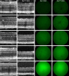
. 2015 Oct;56(11):6961-70.
doi: 10.1167/iovs.15-16969.

In Vivo Visualization of Endoplasmic Reticulum Stress in the Retina Using the ERAI Reporter Mouse

Affiliations

In Vivo Visualization of Endoplasmic Reticulum Stress in the Retina Using the ERAI Reporter Mouse

Marcel V Alavi et al. Invest Ophthalmol Vis Sci. 2015 Oct.

Abstract

Purpose: Endoplasmic reticulum (ER) stress activates inositol requiring enzyme 1 (IRE1), a key regulator of the unfolded protein response. The ER stress activated indicator (ERAI) transgenic mouse expresses a yellow fluorescent GFP variant (Venus) when IRE1 is activated by ER stress. We tested whether ERAI mice would allow for real-time longitudinal studies of ER stress in living mouse eyes.

Methods: We chemically and genetically induced ER stress, and qualitatively and quantitatively studied the Venus signal by fluorescence ophthalmoscopy. We determined retinal cell types that contribute to the signal by immunohistology, and we performed molecular and biochemical assays using whole retinal lysates to assess activity of the IRE1 pathway.

Results: We found qualitative increase in vivo in fluorescence signal at sites of intravitreal tunicamycin injection in ERAI eyes, and quantitative increase in ERAI mice mated to RhoP23H mice expressing ER stress-inducing misfolded rhodopsin protein. As expected, we found that increased Venus signal arose primarily from photoreceptors in RhoP23H/+;ERAI mice. We found increased Xbp1S and XBP1s transcriptional target mRNA levels in RhoP23H/+;ERAI retinas compared to Rho+/+;ERAI retinas, and that Venus signal increased in ERAI retinas as a function of age.

Conclusions: Fluorescence ophthalmoscopy of ERAI mice enables in vivo visualization of retinas undergoing ER stress. ER stress activated indicator mice enable identification of individual retinal cells undergoing ER stress by immunohistochemistry. ER stress activated indicator mice show higher Venus signal at older ages, likely arising from amplification of basal retinal ER stress levels by GFP's inherent stability.


Figures
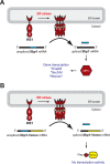
Figure 1
Figure 1
Schematic of the mammalian IRE1 pathway and the function of the XBP1-Venus reporter. Unfolded proteins in the ER (= ER stress) activate IRE1, which splices out an intron of the Xbp1 mRNA. Spliced Xbp1 encodes the transcription factor XBP1s, which upregulates proteins that alleviate ER stress (A). Upon activation, IRE1 also can remove an intron of an Xbp1-Venus reporter transgene in ERAI mice. Spliced Xbp1-Venus mRNA encodes a transcriptional inactive, cytosolic XBP1-Venus fusion protein, which allows for monitoring IRE1 activity by its fluorescence signal (B).
Figure 2
Figure 2
In vivo monitoring IRE1 activation upon tunicamycin injections in ERAI mice. Dimethyl sulfoxide–injected eyes showed no pathologic findings after 2 days (AC), while tunicamycin-injected eyes showed disbanding of the photoreceptor segments (asterisk in [E]) and increased fluorescence at the injection site ([F], superior hemisphere). With progressing degeneration fluorescence signals extended beyond the superior hemisphere (GI) until most of the outer retinal layers were missing and the superior hemisphere showed little or no fluorescence anymore (KL). PI, post injection; GCL, ganglion cell layer; IPL, inner plexiform layer; INL, inner nuclear layer; OPL, outer plexiform layer; ONL, outer nuclear layer; OS/RPE, outer segments–RPE complex.
Figure 3
Figure 3
In vivo assessment of genetically-induced ER stress in retinas of RhoP23H/+;ERAI mice. Misfolded rhodopsin causes ER stress and progressive photoreceptor degeneration. Nontransgenic wild-type mice (AC) did not show any detectable fundus fluorescence in the Venus wavelength range, while Rho+/+;ERAI mice (DF) and RhoP23H/+;ERAI mice (GH) showed clearly detectable Venus fluorescence signals, which were highly significantly stronger in RhoP23H/+;ERAI mice (I). ***P < 0.001.
Figure 4
Figure 4
Age-dependent increase in fluorescence in Rho+/+;ERAI mice and RhoP23H/+;ERAI mice. Compared to Rho+/+;ERAI mice (AE), we found progressive photoreceptor degeneration in RhoP23H/+;ERAI mice (FJ). Fluorescence ophthalmoscopy revealed age-dependent increase in Venus fluorescence in Rho+/+;ERAI (KO) and RhoP23H/+;ERAI (PT) mouse eyes, which appeared stronger in RhoP23H/+;ERAI than in Rho+/+;ERAI eyes at younger ages.
Figure 5
Figure 5
Immunohistochemistry against Venus and rhodopsin in Rho+/+;ERAI and RhoP23H/+;ERAI mice. Rho+/+;ERAI eyes (A, B) and RhoP23H/+;ERAI eyes (C) showed Venus labeling mainly in the photoreceptor layers, as well as single cells in the INL and GCL. Note the increased Venus labeling from P30 to P120 in Rho+/+;ERAI mice (A, B). Rhodopsin (red), Venus (by anti-GFP, green), nuclear stain (DAPI, blue). Scale bar: 20 μm.
Figure 6
Figure 6
Evaluation of photoreceptor loss and Venus signals in Rho+/+;ERAI and RhoP23H/+;ERAI mouse retinas. Progressive photoreceptor loss in RhoP23H/+ retinas compared to Rho+/+ retinas (A). Western blot analysis revealed age-dependent increased antibody labeling against Venus and the amino-terminal Flag tag of the ERAI construct compared to Hsp90 and β-tubulin in Rho+/+;ERAI retinas (B). Fundus fluorescence quantification at different ages showed linearly increased Venus in Rho+/+;ERAI mice, while in P270 RhoP23H/+;ERAI mice fluorescence leveled off (C). The Venus signal at younger ages was significantly higher in RhoP23H/+;ERAI mice than in Rho+/+;ERAI mice (C). Taking ONL thickness into account (shown in [A]), the relative fluorescence signals in RhoP23H/+;ERAI mice vastly surpassed signals in Rho+/+;ERAI mice at all ages (D). *P < 0.05, ***P < 0.001.
Figure 7
Figure 7
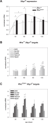
Xbp1S mRNA levels and expression levels of XBP1s downstream targets in Rho+/+ and RhoP23H/+ mouse retinas. RhoP23H/+ retinas showed significantly elevated Xbp1S transcript levels compared to Rho+/+ retinas at ages P30 and P60 (A). Rho+/+ mice also showed a rather small, but significant, age-dependent increase of Xbp1S levels between P30 and P60 in (A). Temporal expression of the XBP1 downstream targets Sec24d, Dnajb9, Herpud1, and Hspa5 followed Xbp1S levels in both genotypes (B, C), while Ddit3, a target of the PERK signaling pathway of the UPR, did not show any changes (B, C). *P < 0.05, ***P < 0.001.

References

    1. Alberts B. Molecular Biology of the Cell. 5th ed. New York, NY: Garland Science; 2008: 1 v. (various pages).
    1. Walter P,, Ron D. The unfolded protein response: from stress pathway to homeostatic regulation. Science. 2011. ; 334: 1081–1086. - PubMed
    1. Zhang SX,, Sanders E,, Fliesler SJ,, Wang JJ. Endoplasmic reticulum stress and the unfolded protein responses in retinal degeneration. Exp Eye Res. 2014; 125: 30–40. - PMC - PubMed
    1. Zode GS,, Sharma AB,, Lin X,, et al. Ocular-specific ER stress reduction rescues glaucoma in murine glucocorticoid-induced glaucoma. J Clin Invest. 2014; 124: 1956–1965. - PMC - PubMed
    1. Zode GS,, Kuehn MH,, Nishimura DY,, et al. Reduction of ER stress via a chemical chaperone prevents disease phenotypes in a mouse model of primary open angle glaucoma. J Clin Invest. 2011. ; 121: 3542–3553. - PMC - PubMed

Publication types

MeSH terms