

Lung Pathology in U.S. Coal Workers with Rapidly Progressive Pneumoconiosis Implicates Silica and Silicates
- PMID: 26513613
- PMCID: PMC4824937
- DOI: 10.1164/rccm.201505-1014OC
Lung Pathology in U.S. Coal Workers with Rapidly Progressive Pneumoconiosis Implicates Silica and Silicates
Abstract
Rationale: Recent reports of progressive massive fibrosis and rapidly progressive pneumoconiosis in U.S. coal miners have raised concerns about excessive exposures to coal mine dust, despite reports of declining dust levels.
Objectives: To evaluate the histologic abnormalities and retained dust particles in available coal miner lung pathology specimens, and to compare these findings with those derived from corresponding chest radiographs.
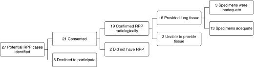
Methods: Miners with severe disease and available lung tissue were identified through investigator outreach. Demographic as well as smoking and work history information was obtained. Chest radiographs were interpreted according to the International Labor Organization classification scheme to determine if criteria for rapidly progressive pneumoconiosis were confirmed. Pathology slides were scored by three expert pulmonary pathologists using a standardized nomenclature and scoring system.
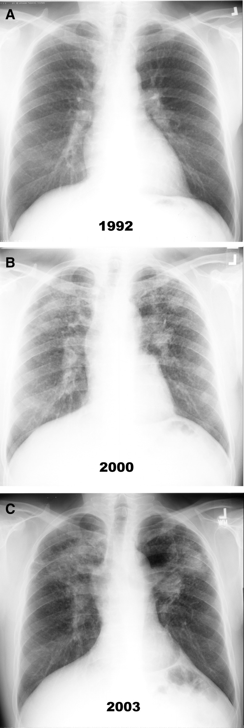
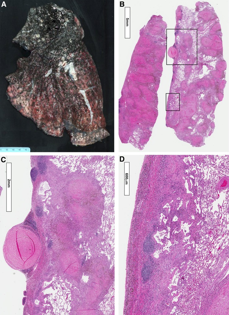
Measurements and main results: Thirteen cases were reviewed, many of which had features of accelerated silicosis and mixed dust lesions. Twelve had progressive massive fibrosis, and 11 had silicosis. Only four had classic lesions of simple coal workers' pneumoconiosis. Four had diffuse interstitial fibrosis with chronic inflammation, and two had focal alveolar proteinosis. Polarized light microscopy revealed large amounts of birefringent mineral dust particles consistent with silica and silicates; carbonaceous coal dust was less prominent. On the basis of chest imaging studies, specimens with features of silicosis were significantly associated (P = 0.047) with rounded (type p, q, or r) opacities, whereas grade 3 interstitial fibrosis was associated (P = 0.02) with the presence of irregular (type s, t, or u) opacities.
Conclusions: Our findings suggest that rapidly progressive pneumoconiosis in these miners was associated with exposure to coal mine dust containing high concentrations of respirable silica and silicates.
Keywords: anthracosis; coal mining; pathology; pneumoconiosis; silicosis.
Figures
Comment in
-
Pneumoconiosis Redux. Coal Workers' Pneumoconiosis and Silicosis Are Still a Problem.Am J Respir Crit Care Med. 2016 Mar 15;193(6):603-5. doi: 10.1164/rccm.201511-2154ED. Am J Respir Crit Care Med. 2016. PMID: 26977968 No abstract available.
-
Down Under in the Coal Mines.Am J Respir Crit Care Med. 2016 Sep 15;194(6):772-3. doi: 10.1164/rccm.201603-0615LE. Am J Respir Crit Care Med. 2016. PMID: 27628081 No abstract available.
-
Reply: Coal Mine Dust Lung Disease That Persists below the Surface of Surveillance: Down Under.Am J Respir Crit Care Med. 2016 Sep 15;194(6):773-4. doi: 10.1164/rccm.201604-0779LE. Am J Respir Crit Care Med. 2016. PMID: 27628082 No abstract available.
References
-
- Federal Coal Mine Health and Safety Act of 1969, Public Law 91-173, 30 U.S.C. ch. 22, §§ 801 et seq.
-
- International Labour Office. Geneva, Switzerland: International Labour Office; 2002. Guidelines for the use of the ILO international classification of radiographs of pneumoconioses.
-
- National Institute for Occupational Safety and Health, Centers for Disease Control and Prevention. Work-related lung disease (WoRLD) surveillance report [updated 2008 Jun 23; accessed 2015 Nov 11]. Available from: http://www2a.cdc.gov/drds/worldreportdata/tocArchive.asp.
-
- Antao V, Petsonk EL, Attfield MD Centers for Disease Control and Prevention (CDC) Advanced cases of coal workers’ pneumoconiosis—two counties, Virginia, 2006. MMWR Morb Mortal Wkly Rep. 2006;55:909–913. - PubMed
-
- Wade WA, Petsonk EL, Young B, Mogri I. Severe occupational pneumoconiosis among West Virginian coal miners: one hundred thirty-eight cases of progressive massive fibrosis compensated between 2000 and 2009. Chest. 2011;139:1458–1462. - PubMed
Publication types
MeSH terms
Substances
Grants and funding
LinkOut - more resources
Full Text Sources
Other Literature Sources
