. 2016 Jan;45(1):88-97.
doi: 10.1097/SHK.0000000000000478.

AP39, A Mitochondrially Targeted Hydrogen Sulfide Donor, Exerts Protective Effects in Renal Epithelial Cells Subjected to Oxidative Stress in Vitro and in Acute Renal Injury in Vivo

Affiliations

AP39, A Mitochondrially Targeted Hydrogen Sulfide Donor, Exerts Protective Effects in Renal Epithelial Cells Subjected to Oxidative Stress in Vitro and in Acute Renal Injury in Vivo

Akbar Ahmad et al. Shock. 2016 Jan.

Abstract

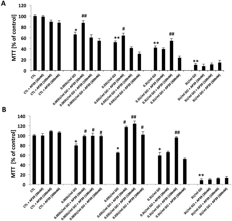
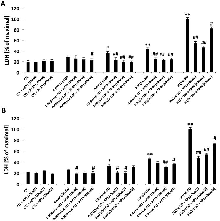
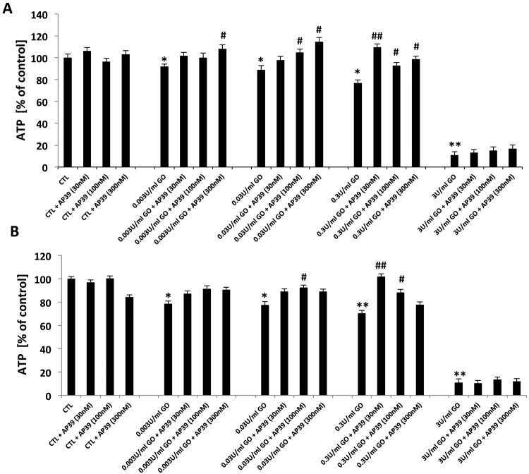
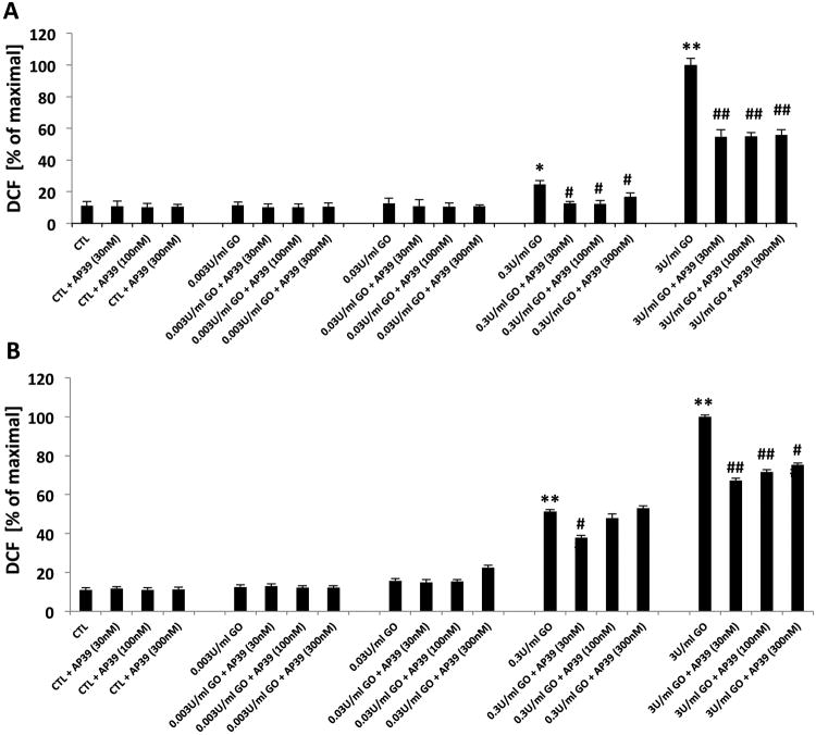
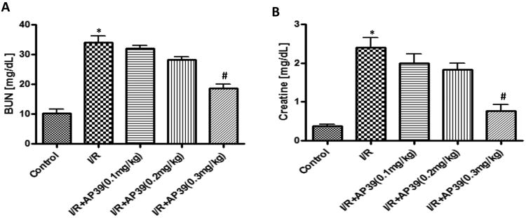
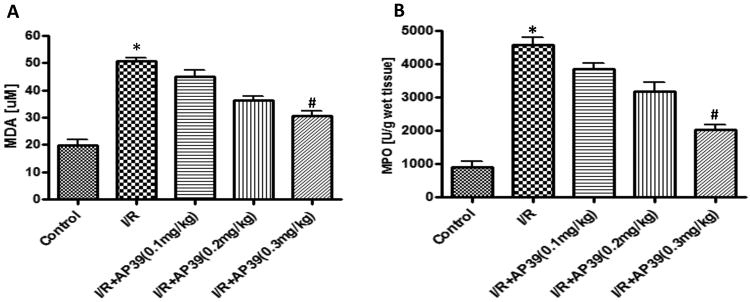
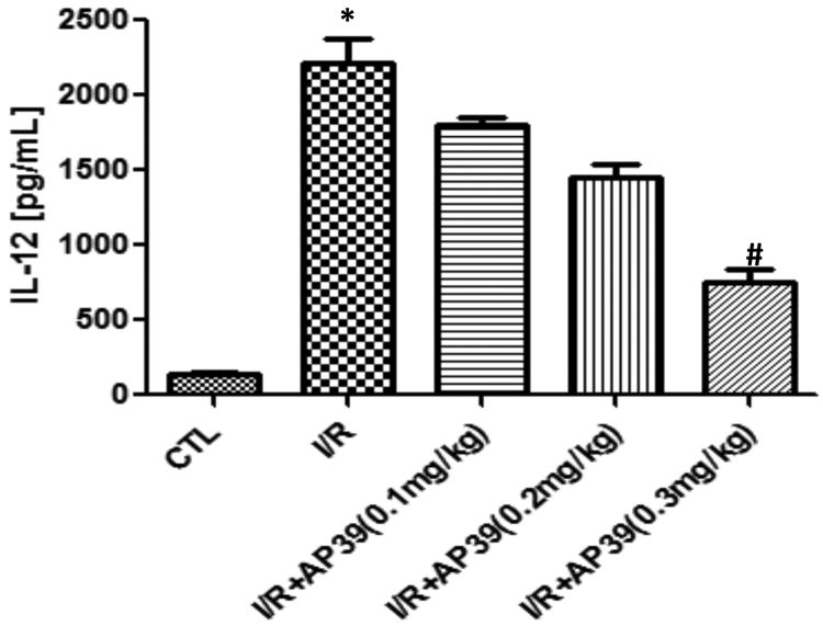
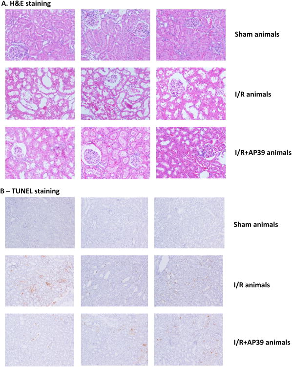
This study evaluated the effects of AP39 [(10-oxo-10-(4-(3-thioxo-3H-1,2-dithiol-5yl) phenoxy)decyl) triphenyl phosphonium bromide], a mitochondrially targeted donor of hydrogen sulfide (H2S) in an in vitro model of hypoxia/oxidative stress injury in NRK-49F rat kidney epithelial cells (NRK cells) and in a rat model of renal ischemia-reperfusion injury. Renal oxidative stress was induced by the addition of glucose oxidase, which generates hydrogen peroxide in the culture medium at a constant rate. Glucose oxidase (GOx)-induced oxidative stress led to mitochondrial dysfunction, decreased intracellular ATP content, and, at higher concentrations, increased intracellular oxidant formation (estimated by the fluorescent probe 2, 7-dichlorofluorescein, DCF) and promoted necrosis (estimated by the measurement of lactate dehydrogenase release into the medium) of the NRK cells in vitro. Pretreatment with AP39 (30-300 nM) exerted a concentration-dependent protective effect against all of the above effects of GOx. Most of the effects of AP39 followed a bell-shaped concentration-response curve; at the highest concentration of GOx tested, AP39 was no longer able to afford cytoprotective effects. Rats subjected to renal ischemia/reperfusion responded with a marked increase (over four-fold over sham control baseline) blood urea nitrogen and creatinine levels in blood, indicative of significant renal damage. This was associated with increased neutrophil infiltration into the kidneys (assessed by the myeloperoxidase assay in kidney homogenates), increased oxidative stress (assessed by the malondialdehyde assay in kidney homogenates), and an increase in plasma levels of IL-12. Pretreatment with AP39 (0.1, 0.2, and 0.3 mg/kg) provided a dose-dependent protection against these pathophysiological alterations; the most pronounced protective effect was observed at the 0.3 mg/kg dose of the H2S donor; nevertheless, AP39 failed to achieve a complete normalization of any of the injury markers measured. The partial protective effects of AP39 correlated with a partial improvement of kidney histological scores and reduced TUNEL staining (an indicator of DNA damage and apoptosis). In summary, the mitochondria-targeted H2S donor AP39 exerted dose-dependent protective effects against renal epithelial cell injury in vitro and renal ischemia-reperfusion injury in vivo. We hypothesize that the beneficial actions of AP39 are related to the reduction of cellular oxidative stress, and subsequent attenuation of various positive feed-forward cycles of inflammatory and oxidative processes.


Conflict of interest statement

Competing interests: M.W., M.E.W. and the University of Exeter have intellectual property (patent filings) related to AP39 and its therapeutic uses. The other authors declare no conflicts of interest in relationship to this study.

Figures

Figure 1
Figure 1. Effect of AP39 on the viability of oxidatively stressed NRK cells
AP39 (30 nM, 100 nM or 300 nM) pretreatment enhances cellular recovery after 1h (A) & 24h (B) of GOx exposure (0.003, 0.03, 0.3 or 3U/ml) on NRK cells, followed by the measurement of MTT conversion. There is a decrease in MTT conversion in GOx treated cells; these effects are attenuated by AP39. Data (% of vehicle-treated control values) are shown as mean ± SEM values from three independent experiments with 4–8 replicates for each end-point. * p<0.05, ** p<0.01 shows a significant decrease in MTT in response to GOx treatment (in the GOx treated group that also received AP39 vehicle, compared to the to baseline control in the absence of GOx or AP39). # p<0.05, ## p<0.01 shows a significant enhancement of MTT by AP39, when compared to its vehicle control in the presence of the same concentration of GOx.
Figure 2
Figure 2. Effect of AP39 on LDH release of oxidatively stressed NRK cells
Cells were exposed to glucose oxidase (0.003, 0.03, 0.3 or 3U/ml) for 1h (A), in another set exposed to 1h followed by a washout and replacement of the medium with fresh tissue culture medium and incubated for a subsequent 24h (B), followed by the measurement of LDH conversion. There is an increase in LDH content of the medium of GOx treated cells; these effects are attenuated by AP39 (30 nM, 100 nM and 300 nM). Data are shown as mean ± SEM values from three independent experiments with 4–8 replicates for each end-point; values are expressed as the maximum LDH release observed at 3 U/ml GOx. * p<0.05, ** p<0.01 shows a significant increase in LDH release in response to GOx treatment (in the GOx treated group that also received AP39 vehicle, compared to the to baseline control in the absence of GOx or AP39). # p<0.05, ## p<0.01 shows a significant reduction of LDH by AP39, when compared to its vehicle control at the same concentration of GOx.
Figure 3
Figure 3. Effect of AP39 on cellular ATP levels of oxidatively stressed NRK cells
NRK cells were subjected to 1h (A) and 24h (B) GOx exposure (0.003, 0.03, 0.3& 3U/ml). Cells were pretreated with AP39 (30 nM, 100 nM or 300 nM) 30 min prior to GOx exposure. AP39-mediated protection was observed at intermediate concentrations of GOx. Data (% of vehicle-treated control values) are shown as mean ± SEM values from three independent experiments with 4–8 replicates for each end-point. * p<0.05, ** p<0.01 shows a significant decrease in ATP levels in response to GOx treatment, (in the GOx treated group that also received AP39 vehicle, compared to the to baseline control in the absence of GOx or AP39). # p<0.05, ## p<0.01 shows a significant enhancement of ATP levels by AP39, when compared to its corresponding control at the same concentration of GOx.
Figure 4
Figure 4. Effect of AP39 on DCF production of oxidatively stressed NRK cells
Increase in DCF fluorescence in response to exposure of GOx for 1h (A) and 24h (B) incubation in tissue culture medium of NRK cells is shown. Pretreatment with AP39 30 min prior to GOx exposure observed concentration dependent protective effect. Data are shown as mean ± SEM values from three independent experiments with 4–8 replicates for each end-point; values are expressed as the maximum DCF production observed at 3 U/ml GOx. * p<0.05, ** p<0.01 shows a significant increase in DCF fluorescence in response to GOx treatment, when compared to baseline control (in the absence of GOx or AP39). # p<0.05, ## p<0.01 shows a significant reduction of DCF fluorescence by AP39, when compared to its corresponding control at the same concentration of GOx.
Figure 5
Figure 5. Effect of AP39 on BUN and creatinine plasma levels in a renal ischemia-reperfusion model in the rat
Renal I/R significantly impaired glomerular function, as evidenced by markedly increased (over 4-fold over sham control baseline) blood urea nitrogen (A) and creatinine levels (B); this increase was concentration-dependently reduced by AP39; most pronouncedly at the 0.3 mg/kg dose of the H2S donor. Data are shown as mean ± SEM of 10 animals; *p<0.05 show significant increases in the vehicle-treated I/R group, compared to the sham control; # p<0.05 shows a significant protective effect of AP39 in I/R compared to the I/R group treated with AP39 vehicle.
Figure 6
Figure 6. Effect of AP39 on MPO and MDA levels in a renal ischemia-reperfusion model in the rat
Renal I/R significantly increased renal MPO (A) and renal MDA levels (B); this increase was concentration-dependently reduced by AP39; most pronouncedly at the 0.3 mg/kg dose of the H2S donor. Data are shown as mean ± SEM of 10 animals; *p<0.05 show significant increases in the vehicle-treated I/R group, compared to the sham control; # p<0.05 shows a significant protective effect of AP39 in I/R compared to the I/R group treated with AP39 vehicle.
Figure 7
Figure 7. Effect of AP39 on plasma IL-12 levels in a renal ischemia-reperfusion model in the rat
Renal I/R significantly increased plasma IL-12 levels; this increase was concentration-dependently reduced by AP39; most pronouncedly at the 0.3 mg/kg dose of the H2S donor. Data are shown as mean ± SEM of 10 animals; *p<0.05 show significant increases in the vehicle-treated I/R group, compared to the sham control; # p<0.05 shows a significant protective effect of AP39 in I/R compared to the I/R group treated with AP39 vehicle. Data are shown as mean ± SEM of 10 animals.
Figure 8
Figure 8. Effect of AP39 on histopathology and apoptosis in a renal ischemia-reperfusion model in the rat
Hematoxylin and eosin–stained sections are from the outer medulla are shown in (A); TUNEL staining is shown in (B). Top figures represent representative sham controls; middle figures represent vehicle-treated rats subjected to renal I/R; bottom figures represent an AP39 (0.3 mg/kg) treated rat subjected to renal I/R. Please note that neutrophil accumulation within the interstitium of the kidney, as well as the intensity of TUNEL staining was significantly reduced by AP39, but neither the histological damage, nor the TUNEL staining was completely normalized by AP39 treatment. Each group shows 3 representative animals of n=10 animals.

References

    1. Paller MS. The cell biology of reperfusion injury in the kidney. J Invest Med. 1994;42:632–639. - PubMed
    1. Kribben A, Edelstein CL, Schrier RW. Pathophysiology of acute renal failure. J Nephrol. 1999;12:s142–s151. - PubMed
    1. Szabo C. Hydrogen sulphide and its therapeutic potential. Nat Rev Drug Discov. 2007;6:917–935. - PubMed
    1. Wallace JL, Wang R. Hydrogen sulfide-based therapeutics: exploiting a unique but ubiquitous gasotransmitter. Nat Rev Drug Discov. 2015;14:329–345. - PubMed
    1. Szabo C. Medicinal chemistry and therapeutic applications of the gasotransmitters nitric oxide, carbon monoxide and hydrogen sulfide. Burger's Medicinal Chemistry. 2010;5:265–367.

Publication types

MeSH terms