Randomized Controlled Trial
. 2016 May;41(6):1659-69.
doi: 10.1038/npp.2015.336. Epub 2015 Oct 30.

Brain Mapping-Based Model of Δ(9)-Tetrahydrocannabinol Effects on Connectivity in the Pain Matrix

Affiliations
Randomized Controlled Trial

Brain Mapping-Based Model of Δ(9)-Tetrahydrocannabinol Effects on Connectivity in the Pain Matrix

Carmen Walter et al. Neuropsychopharmacology. 2016 May.

Abstract

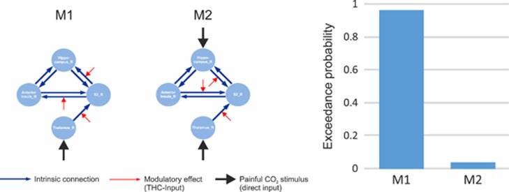
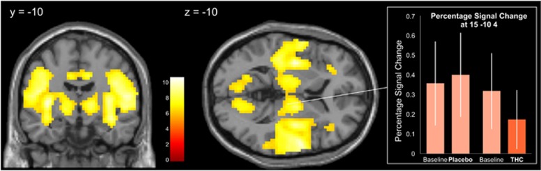
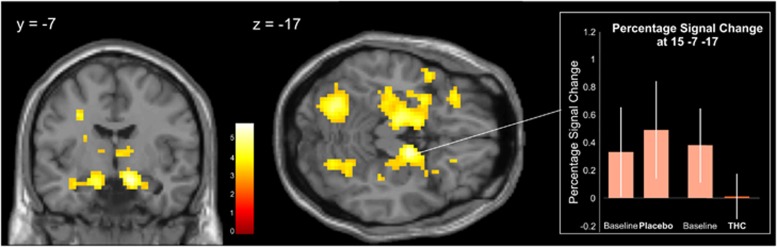
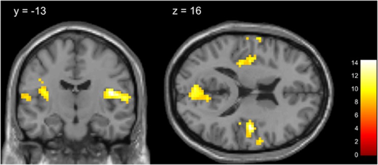
Cannabinoids receive increasing interest as analgesic treatments. However, the clinical use of Δ(9)-tetrahydrocannabinol (Δ(9)-THC) has progressed with justified caution, which also owes to the incomplete mechanistic understanding of its analgesic effects, in particular its interference with the processing of sensory or affective components of pain. The present placebo-controlled crossover study therefore focused on the effects of 20 mg oral THC on the connectivity between brain areas of the pain matrix following experimental stimulation of trigeminal nocisensors in 15 non-addicted healthy volunteers. A general linear model (GLM) analysis identified reduced activations in the hippocampus and the anterior insula following THC administration. However, assessment of psychophysiological interaction (PPI) revealed that the effects of THC first consisted in a weakening of the interaction between the thalamus and the secondary somatosensory cortex (S2). From there, dynamic causal modeling (DCM) was employed to infer that THC attenuated the connections to the hippocampus and to the anterior insula, suggesting that the reduced activations in these regions are secondary to a reduction of the connectivity from somatosensory regions by THC. These findings may have consequences for the way THC effects are currently interpreted: as cannabinoids are increasingly considered in pain treatment, present results provide relevant information about how THC interferes with the affective component of pain. Specifically, the present experiment suggests that THC does not selectively affect limbic regions, but rather interferes with sensory processing which in turn reduces sensory-limbic connectivity, leading to deactivation of affective regions.


Figures

Figure 1
Figure 1
Left: Schematic structure of the two models compared in this study. In all models, seven intrinsic connections were defined (blue lines with arrows indicating the direction of connection). The driving input pain (black lines) enters the model via thalamus (model 1) and via thalamus and hippocampus (model 2), respectively. THC modulates the connection between thalamus and S2 (as known from the PPI analysis) and the connections from S2 to the anterior insula and to the hippocampus (model 1) or reverse, ie, from hippocampus and anterior insula to S2 (Model 2). Right: Result of Bayesian model selection on model level shows that exceedance probability of model 1 (EP: 0.96) exceeds model 2 by far (EP: 0.04).
Figure 2
Figure 2
Brain regions that were activated by the CO2 pain stimulus (main effect ‘stimulus'). The topographies of differences in brain activations are superimposed upon slices of the canonical MR template implemented in SPM8. The significance at voxel level is color coded from red to white with increasing t-values. Voxels are shown at a threshold of p<0.001 (FWE-corrected, t>6.18). The bars show the effect size (mean and standard deviation) at coordinates of the right thalamus used as a seed region for subsequent PPI.
Figure 3
Figure 3
Brain regions that were deactivated by THC administration (interaction ‘drug', ie, placebo or THC) by ‘measurement' (ie, baseline or post-drug session). The topographies of differences in brain activations are superimposed upon slices of the canonical MR template implemented in SPM8. The significance at voxel level is color coded from red to white with increasing t-values. Voxels are shown at a threshold of p<0.001 (uncorrected, t>3.25). The bars show the effect size (mean and standard deviation) at the coordinates of the right hippocampus used as seed region for subsequent PPI.
Figure 4
Figure 4
Brain regions that showed reduced functional connectivity to the seed region right thalamus (x=15, y=−10, z=4) after THC administration (PPI, seed region × (non-THC condition−THC condition)). The topographies of differences in brain activations are superimposed upon slices of the canonical MR template implemented in SPM8. The significance at voxel level is color coded from red to white with increasing t-values. Voxels are shown at a threshold of p<0.05 (FWE-corrected, t>7.94).

References

    1. Ab Aziz CB, Ahmad AH (2006). The role of the thalamus in modulating pain. Malays J Med Sci 13: 11–18. - PMC - PubMed
    1. Abrams DI, Jay CA, Shade SB, Vizoso H, Reda H, Press S et al (2007). Cannabis in painful HIV-associated sensory neuropathy: a randomized placebo-controlled trial. Neurology 68: 515–521. - PubMed
    1. Agarwal N, Pacher P, Tegeder I, Amaya F, Constantin CE, Brenner GJ et al (2007). Cannabinoids mediate analgesia largely via peripheral type 1 cannabinoid receptors in nociceptors. Nat Neurosci 10: 870–879. - PMC - PubMed
    1. Andersson JL, Hutton C, Ashburner J, Turner R, Friston K (2001). Modeling geometric deformations in EPI time series. Neuroimage 13: 903–919. - PubMed
    1. Apkarian AV, Bushnell MC, Treede RD, Zubieta JK (2005). Human brain mechanisms of pain perception and regulation in health and disease. Eur J Pain 9: 463–484. - PubMed

Publication types

MeSH terms