. 2016 Jan 29;291(5):2499-509.
doi: 10.1074/jbc.M115.668236. Epub 2015 Oct 29.

Mechanisms of Calmodulin Regulation of Different Isoforms of Kv7.4 K+ Channels

Affiliations

Mechanisms of Calmodulin Regulation of Different Isoforms of Kv7.4 K+ Channels

Choong-Ryoul Sihn et al. J Biol Chem. .

Abstract

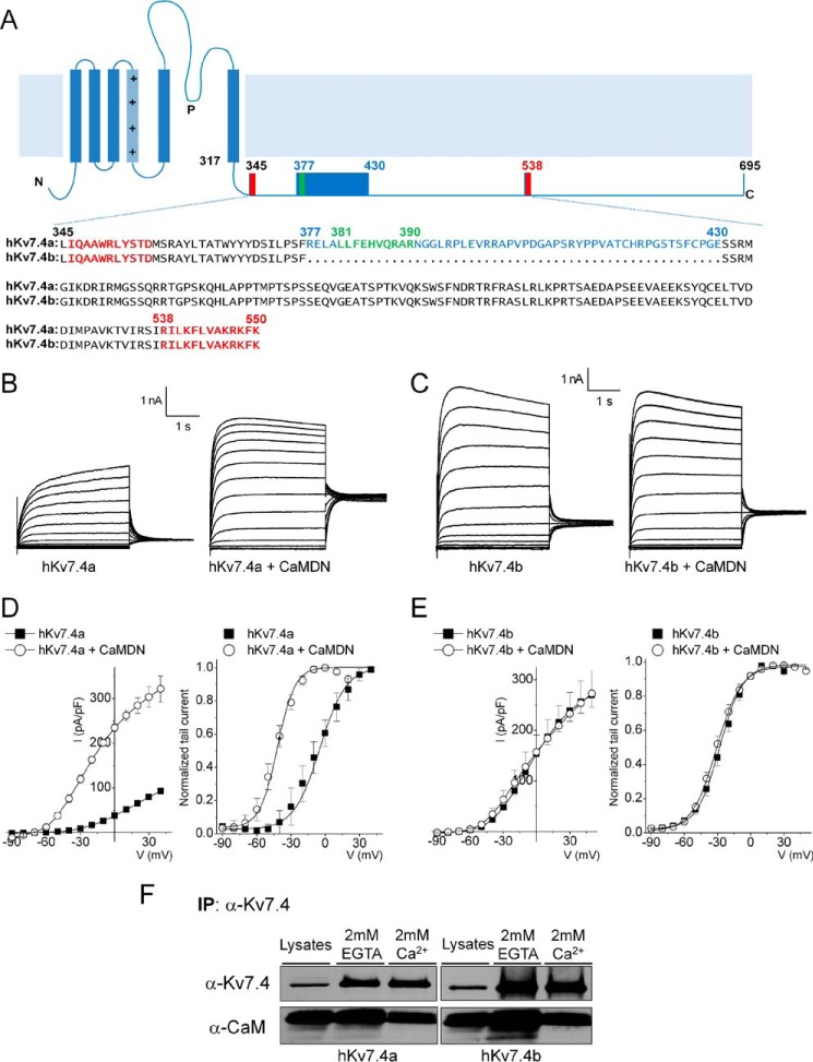
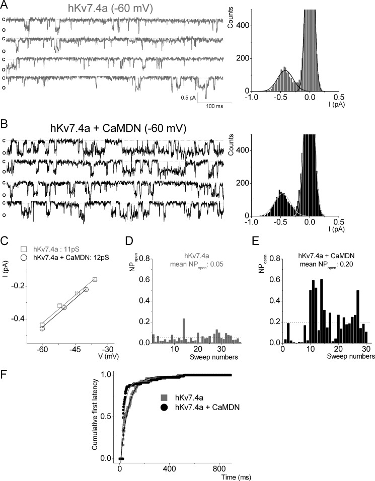
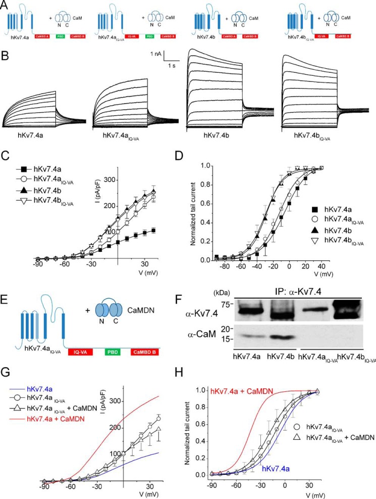
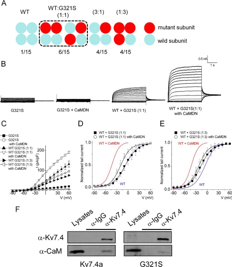
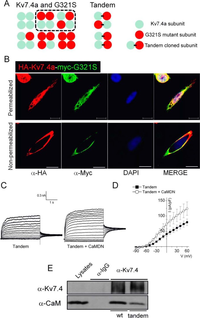
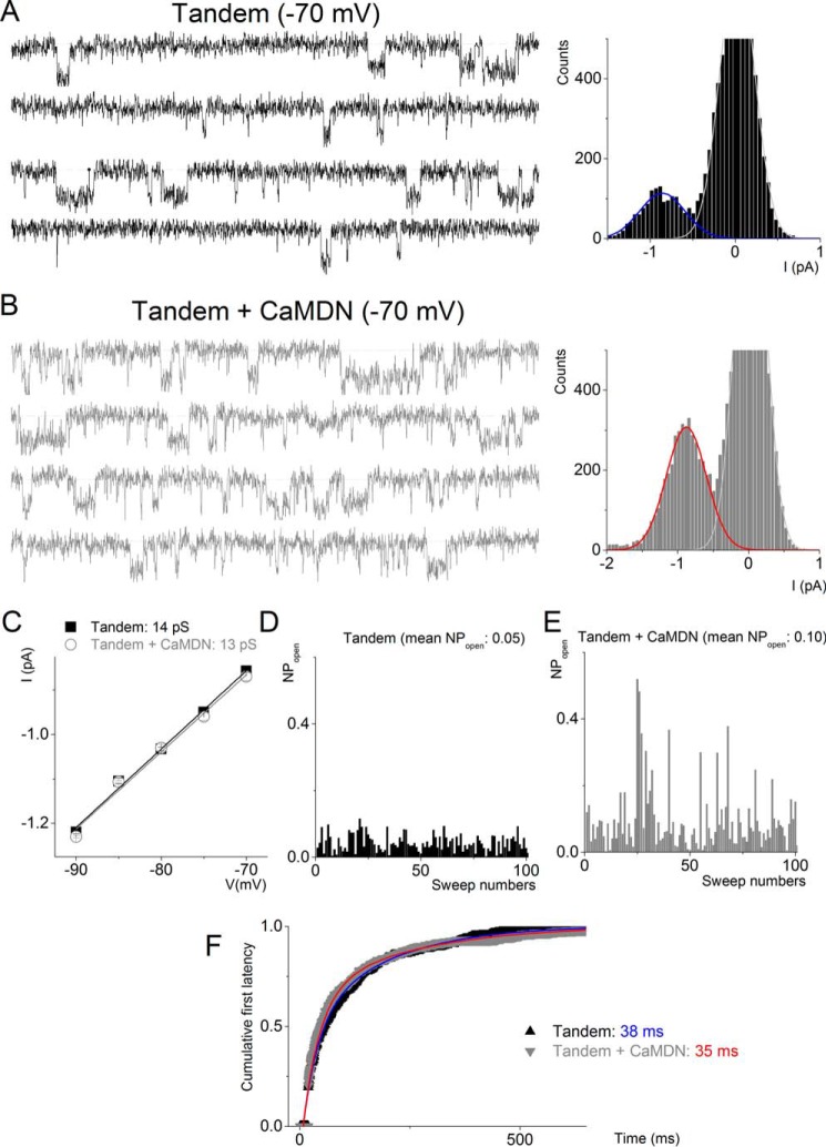
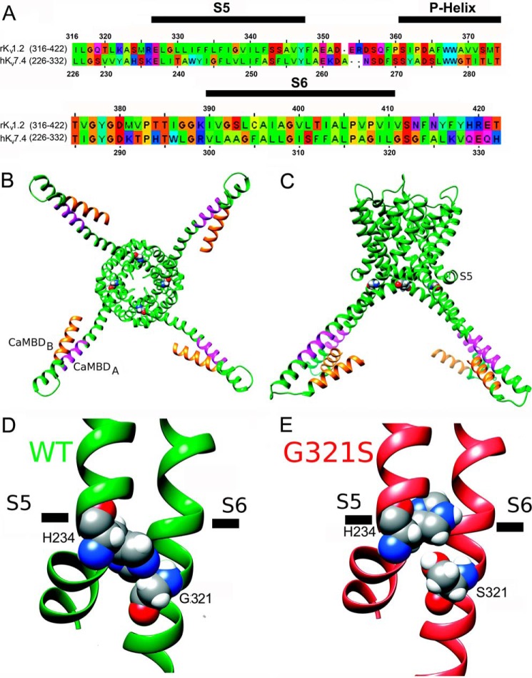
Calmodulin (CaM), a Ca(2+)-sensing protein, is constitutively bound to IQ domains of the C termini of human Kv7 (hKv7, KCNQ) channels to mediate Ca(2+)-dependent reduction of Kv7 currents. However, the mechanism remains unclear. We report that CaM binds to two isoforms of the hKv7.4 channel in a Ca(2+)-independent manner but that only the long isoform (hKv7.4a) is regulated by Ca(2+)/CaM. Ca(2+)/CaM mediate reduction of the hKv7.4a channel by decreasing the channel open probability and altering activation kinetics. We took advantage of a known missense mutation (G321S) that has been linked to progressive hearing loss to further examine the inhibitory effects of Ca(2+)/CaM on the Kv7.4 channel. Using multidisciplinary techniques, we demonstrate that the G321S mutation may destabilize CaM binding, leading to a decrease in the inhibitory effects of Ca(2+) on the channels. Our study utilizes an expression system to dissect the biophysical properties of the WT and mutant Kv7.4 channels. This report provides mechanistic insights into the critical roles of Ca(2+)/CaM regulation of the Kv7.4 channel under physiological and pathological conditions.

Keywords: calcium; calmodulin (CaM); hearing; ion channel; potassium channel.


Figures

FIGURE 1.
FIGURE 1.
The CaMBD linker is required for the Ca2+/CaM-dependent inhibition of the Kv7.4 current. A, schematic of the Kv7.4 α-subunit structure. The C-terminal sequence alignment of the two isoforms (hKv7.4a, NP_004691; hKv7.4b, NP_751895) is shown. Two known CaM binding domains (CaMBDA and CaMBDB) are marked in red, and a putative CaMBD is marked in green. Spliced alteration of hKv7.4 (hKv7.4a versus hKv7.4b) is indicated in blue. B and C, representative traces showing whole-cell recordings from CHO cells transfected with hKv7.4a and hKv7.4b and co-transfected with either empty vector or CaMDN. The current traces were recorded from a holding potential of −70 mV to step potentials ranging from −90 to 40 mV using voltage increments of 10 mV. Tail currents were recorded at −30 mV. D and E, plots of current density (picoampere/picofarad)-voltage relations of currents (left) were derived from CHO cells expressing hKv7.4a (or hKv7.4b) (closed squares, n = 21) and hKv7.4a (or hKv7.4b) + CaMDN (open circles, n = 21). Normalized tail currents of hKv7.4a and hKv7.4a + CaMDN are plotted against the applied voltage to generate the activation curves (right), which were fitted with a Boltzmann function. The half-activation voltages (V½, in millivolt) and slope factor, k, for hKv7.4a were as follows: −5.7 ± 1.1 and 14.1 ± 0.9 (n = 17); for hKv7.4a + CaMDN they were −42.0 ± 1.8 and 11.4 + 1.5 (n = 19), respectively. The V½ and k values for hKv7.4b were as follows: −28.0 ± 2.6 and 10.5 ± 1.4; and for hKv7.4b + CaMDN they were −31.0 ± 1.4 and 11.1 ± 1.0 (n = 21). F, CHO cells were expressed with either hKv7.4a or hKv7.4b for 24 h. After preparation of cell lysates, immunoprecipitation (IP) was accomplished using hKv7.4 antibody with a buffer containing either 2 mm of CaCl2 or EGTA. Association of hKv7.4 with CaM is Ca2+-independent.
FIGURE 2.
FIGURE 2.
CaMDN increases the open probability but not unitary conductance of the Kv7.4a channel. A and B, a set of single-channel traces was recorded from CHO cells expressing either hKv7.4a alone (gray) or hKv7.4a + CaMDN (black) at a −60 mV step potential. The dotted lines represent closed states. Representative amplitude histograms of hKv7.4a alone and hKv7.4a + CaMDN at −60 mV are shown on the right. c, closed; o, open; I, current. C, data from amplitude histograms of applied voltages were used to generate current-voltage relationships. The single-channel conductances were 11 ± 0.6 pS (n = 9) (hKv7.4a) and 12 ± 0.8 pS (n = 10) (hKv7.4a + CaMDN), respectively. D and E, the open probability of hKv7.4a and hKv7.4a + CaMDN. Mean open probabilities are indicated by the dotted lines. F, exponential fits to the first latency histograms are shown. Time constants of hKv7.4a and hKv7.4a + CaMDN are indicated in milliseconds, and their median values are 46 ± 6 and 87 ± 9 ms (n = 7), respectively.
FIGURE 3.
FIGURE 3.
The IQ motif in CaMBDA is necessary but not sufficient for Ca2+/CaM-dependent inhibition of the hKv7.4 current. A, the combinations of hKv7.4 and MT hKv7.4IQ-VA isoforms with endogenous CaM. B, representative current traces of hKv7.4 and hKv7.4IQ-VA recorded from transfected CHO cells. Whole-cell recordings were generated from a holding potential of −70 mV to step potentials ranging from −90 to 40 mV using voltage increments of 10 mV. Tail currents were recorded at −30 mV. C, plots of current density (picoampere/picofarad)-voltage relations were generated from CHO cells expressing hKv7.4 and hKv7.4IQ-VA isoforms (n = 19). I, current. D, normalized tail currents of hKv7.4 (hKv7.4IQ-VA) isoforms were plotted against applied voltages to generate activation curves that were fitted with the Boltzmann function. The V½ (in millivolt) and k values for hKv7.4a and hKv7.4aIQ-VA were as follows: −5.7 ± 1.1 and 14.1 ± 0.9 (n = 17), and −12.2 ± 0.5 and 11.4 ± 0.5, respectively (n = 17). For hKv7.4b and hKv7.4bIQ-VA, the V1/2 and k values were as follows: −28.4 ± 2.0 and 10.1 ± 1.2 (n = 17) and −29.7 ± 1.2 and 10.9 ± 1.0, respectively (n = 17). E, schematic of the experiment performed with hKv7.4aIQ-VA + CaMDN. F, co-immunoprecipitation (IP) experiment showing that association of hKv7.4IQ-VA isoforms with CaM was abolished. G, current density (in picoampere/picofarad)-voltage relations of hKv7.4aIQ-VA alone and hKv7.4aIQ-VA + CaMDN (n = 17). H, normalized tail currents for hKv7.4aIQ-VA were plotted against the applied voltage to generate activation curves fitted with a Boltzmann function. The V½ (in millivolt) and k values for hKv7.4aIQ-VA were as follows: −12.2 ± 0.5 and 11.4 ± 0.5 (n = 17); and for hKv7.4aIQ-VA + CaMDN they were −19.0 ± 0.7 and 11.8 ± 0.7, respectively (n = 17). Blue and red curves denote data from Fig. 1D for comparison with hKv7.4a and hKv7.4 + CaMDN, respectively.
FIGURE 4.
FIGURE 4.
Co-expressing the hKv7.4a G321S MT subunit with the WT subunit reduces current density but attenuates the effect of CaMDN on the activation curve. A, schematic of expected channel combinations with hKv7.4a WT and G321S MT type subunits. The numbers presented at the bottom show the potential membrane-expressed channel populations formed from each different ratio of WT and MT subunit. B, representative traces show the whole-cell recording from CHO cells transfected with hKv7.4a WT and G321S MT subunits in a 1:1 ratio (WT:G321S) with either empty vector or CaMDN. The current traces were recorded from a holding potential of −70 mV to step potentials ranging from −90 to 60 mV using voltage increments of 10 mV. Tail currents were recorded at −30 mV. C, plots of current density (picoampere/picofarad)-voltage relations of currents were derived from CHO cells expressing different combinations of hKv7.4a G321S alone, hKv7.4a + CaMDN, 1:1 or 1:3 ratio of (WT:G321S) alone, or with CaMDN. D and E, normalized tail currents of 1:1 ratio (WT:G321S) alone and 1:1 ratio (WT:G321S) + CaMDN are plotted on each voltage applied and generated the activation curves fitted with a Boltzmann function. Blue and red curves denote data from Fig. 1D for comparison with hKv7.4a and hKv7.4 + CaMDN, respectively. F, CHO cells were expressed with either hKv7.4a or hKv7.4a G321S for 24 h. After preparation of cell lysates, immunoprecipitation was accomplished with Kv7.4 antibody to test CaM binding to the channel subunit.
FIGURE 5.
FIGURE 5.
Tandem channel subunits clarify the effect of the MT subunit. A, schematic of expected channel combinations when cells express the hKv7.4 WT and G321S MT subunit or tandem clones. B, the tandem clone has two epitopes, HA and myc, located at the extracellular loop between the S1 and S2 transmembrane domains of Kv7.4a WT and G321S MT, respectively. Anti-HA and anti-myc antibodies were used for the immunofluorescence assay with the conditions either permeabilized or non-permeabilized. Scale bars = 10 μm. C, representative traces showing the whole-cell recording from CHO cells transfected with tandem clones alone or with CaMDN. The current traces were recorded from a holding potential of −70 mV to step potentials ranging from −90 to 60 mV using voltage increments of 10 mV. Tail currents were recorded at −30 mV. D, plots of current (I) density (picoampere/picofarad)-voltage relations of currents were derived from CHO cells expressing tandem clones with either empty vector or CaMDN. E, CHO cells were expressed with either hKv7.4a or tandem clones for 24 h. After preparation of cell lysates, immunoprecipitation was accomplished with Kv7.4 antibody to test CaM binding to the channel subunit.
FIGURE 6.
FIGURE 6.
CaMDN increases the open probability but not unitary conductance of tandem-cloned channels. A and B, representative sets of single-channel traces were recorded from CHO cells expressed with either Tandem alone (black) or Tandem + CaMDN (gray) at a −70 mV step potential. The dotted lines represent closed states. Amplitude histograms of Tandem + CaMWT and Tandem + CaMDN at −70 mV were fitted with a Gaussian function to find a peak point. I, current. C, data from amplitude histograms of applied voltages were used to generate current-voltage relationships. The single-channel conductances were 13 ± 0.6 pS (n = 8) (Tandem alone) and 13 ± 0.2 (n = 7) pS (Tandem + CaMDN), respectively. D and E, the open probability data were generated from at least 200 sweeps at a specific test potential using recordings from Tandem alone and Tandem + CaMDN. Mean open probabilities are shown at the top. F, exponential fits to the first latency histograms. Time constants of Tandem alone (blue) and Tandem + CaMDN (red) are indicated in milliseconds.
FIGURE 7.
FIGURE 7.
Structural modeling of pore-forming domains and CaMBDs in the hKv7.4 WT and G321S MT. A, sequence alignment of the pore-forming domains of the hKv7.4 and rKv1.2 channels. The S5, P-helix, and S6 regions from the same subunit are marked by black bars above the sequence. B, structural model of the hKv7.4 WT viewed from the intracellular side of the membrane. The CaMBD A and B regions in the C terminus are colored orange and magenta, respectively. Gly-321 is shown in spacefilling representation. C, transmembrane view of the hKv7.4 model shown in B. D, close-up view of the structural model of the hKv7.4 WT. Gly-321 and His-234 are shown in spacefilling representation and labeled. E, close-up view of the structural model of the hKv7.4 G321S MT.

References

    1. Yus-Najera E., Santana-Castro I., and Villarroel A. (2002) The identification and characterization of a noncontinuous calmodulin-binding site in noninactivating voltage-dependent KCNQ potassium channels. J. Biol. Chem. 277, 28545–28553 - PubMed
    1. Mooseker M. S., and Cheney R. E. (1995) Unconventional myosins. Annu. Rev. Cell Dev. Biol. 11, 633–675 - PubMed
    1. Tadross M. R., Dick I. E., and Yue D. T. (2008) Mechanism of local and global Ca2+ sensing by calmodulin in complex with a Ca2+ channel. Cell 133, 1228–1240 - PMC - PubMed
    1. Wingo T. L., Shah V. N., Anderson M. E., Lybrand T. P., Chazin W. J., and Balser J. R. (2004) An EF-hand in the sodium channel couples intracellular calcium to cardiac excitability. Nat. Struct. Mol. Biol. 11, 219–225 - PubMed
    1. Gamper N., and Shapiro M. S. (2003) Calmodulin mediates Ca2+-dependent modulation of M-type K+ channels. J. Gen. Physiol. 122, 17–31 - PMC - PubMed

Publication types

MeSH terms

Associated data

LinkOut - more resources