

Pathophysiology of lipid droplet proteins in liver diseases
- PMID: 26515554
- PMCID: PMC4744586
- DOI: 10.1016/j.yexcr.2015.10.021
Pathophysiology of lipid droplet proteins in liver diseases
Abstract
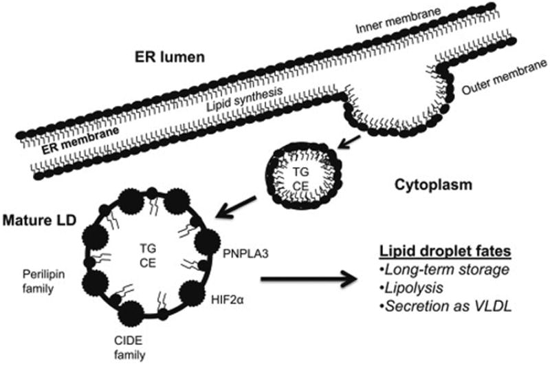
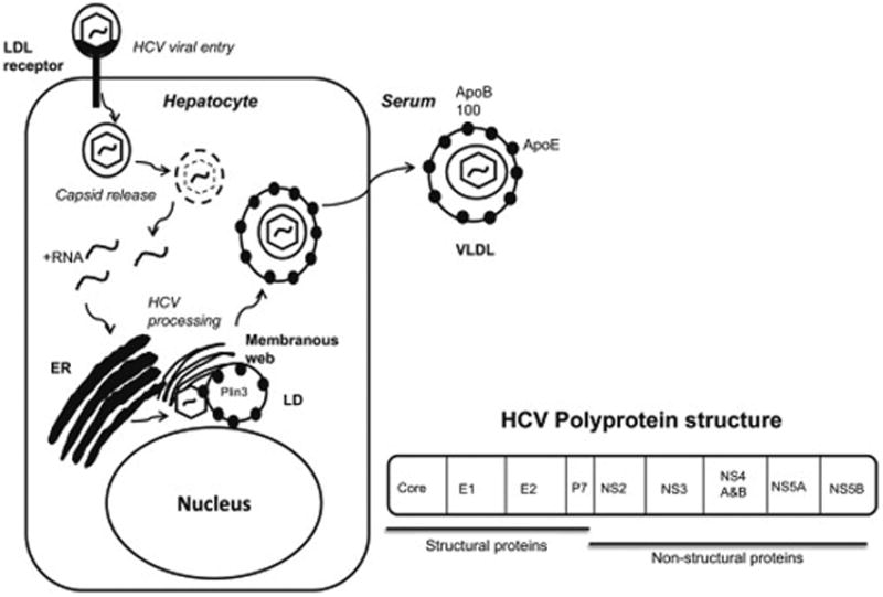
Cytosolic lipid droplets (LDs) are present in most cell types, and consist of a core comprising neutral lipids, mainly triglycerides and sterol esters, surrounded by a monolayer of phospholipids. LDs are heterogeneous in their structure, chemical composition, and tissue distribution. LDs are coated by several proteins, including perilipins and other structural proteins, lipogenic enzymes, lipases and membrane-trafficking proteins. Five proteins of the perilipin (PLIN) family (PLIN1 (perilipin), PLIN2 (adipose differentiation-related protein), PLIN3 (tail-interacting protein of 47kDa), PLIN4 (S3-12), and PLIN5 (myocardial lipid droplet protein)), are associated with LD formation. More recently, the CIDE family of proteins, hypoxia-inducible protein 2 (HIG2), and patanin-like phospholipase domain-containing 3 (PNPLA3) have also gained attention in hepatic LD biology. Evidence suggests that LD proteins are involved in the pathophysiology of fatty liver diseases characterized by excessive lipid accumulation in hepatocytes. This review article will focus on how hepatic LDs and their associated proteins are involved in the pathogenesis of three chronic liver conditions: hepatitis C virus infection, non-alcoholic fatty liver disease, and alcoholic liver disease.
Keywords: Alcohol; Diabetes; HCV; Hepatitis; Lipid droplet; Lipids; Liver; NAFLD; Obesity; Perilipin; Steatosis.
Copyright © 2015 Elsevier Inc. All rights reserved.
Conflict of interest statement
Figures
References
-
- Ploegh HL. A lipid-based model for the creation of an escape hatch from the endoplasmic reticulum. Nature. 2007;448:435–438. - PubMed
-
- Gimm T, Wiese M, Teschemacher B, Deggerich A, Schodel J, Knaup KX, Hackenbeck T, Hellerbrand C, Amann K, Wiesener MS, Honing S, Eckardt KU, Warnecke C. Hypoxia-inducible protein 2 is a novel lipid droplet protein and a specific target gene of hypoxia-inducible factor-1. Faseb J. 2010;24:4443–4458. - PubMed
Publication types
MeSH terms
Substances
Grants and funding
LinkOut - more resources
Full Text Sources
Other Literature Sources
Molecular Biology Databases
Research Materials
Miscellaneous
