. 2015 Dec 15;6(40):42952-62.
doi: 10.18632/oncotarget.5518.

Hepatitis B virus (HBV) receptors: Deficiency in tumor results in scant HBV infection and overexpression in peritumor leads to higher recurrence risk

Affiliations

Hepatitis B virus (HBV) receptors: Deficiency in tumor results in scant HBV infection and overexpression in peritumor leads to higher recurrence risk

Ying-Ying Jing et al. Oncotarget. .

Abstract

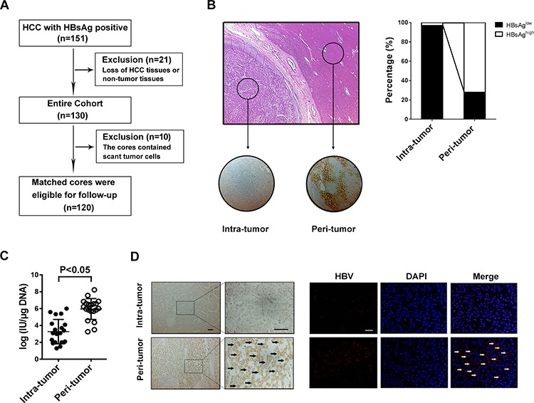
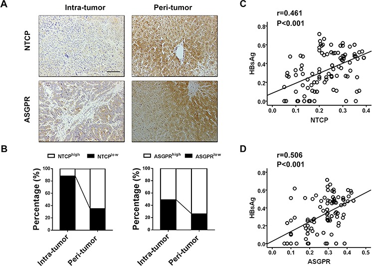
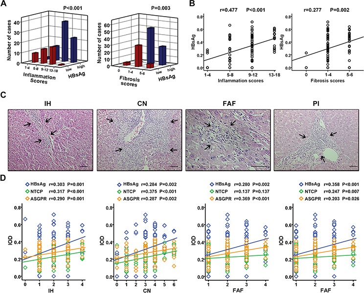
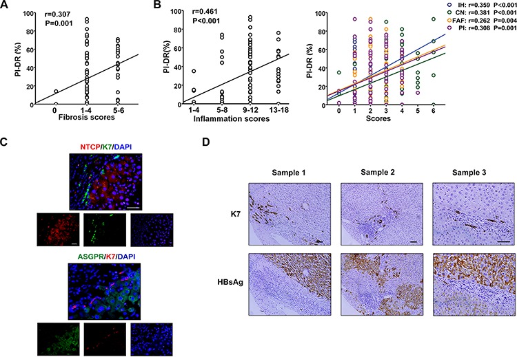
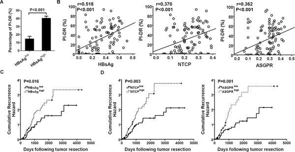
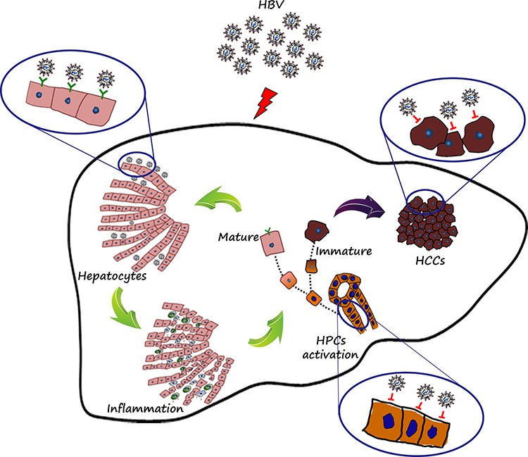
Hepatitis B virus (HBV) infection is a risk factor for hepatocarcinogenesis and recurrence. Here, we sought to characterize intratumoral and peritumoral expression of HBsAg and its specific receptors in HBsAg-positive hepatocellular carcinoma (HCC) patients and further examined their correlation with the recurrence-free survival (RFS). HCC tissue and adjacent normal tissue specimens were acquired from HBsAg-positive patients. The presence of HBsAg and receptors, as well as hepatic progenitor cells (HPCs) were detected by tissue microassay and immunohistochemistry. Necroinflammatory activity was evaluated by HE staining. The mean IOD of HBsAg and HBV DNA in the intratumoral tissues was markedly lower than that in the peritumoral tissues (P < 0.001). Pearson correlation analysis further showed a significant correlation between the expression of HBsAg and NTCP (r = 0.461, P < 0.001) or ASGPR (r = 0.506, P < 0.001) in peritumoral tissues. And the peritumoral HBsAg and receptors presented a positive association with necroinflammatory activity (P < 0.05). Inflammation induced by HBV infection presented a positive association with HPCs activation (P < 0.05). Additionally, due to lack of HBV receptors, HPCs was not preferentially infected with HBV, but activated HPCs had a significant correlation with HBsAg expression in peritumoral tissues, and the peritumoral HPCs activation was associated with RFS of HCC patients, therefore, the overexpression of HBsAg and receptors in peritumor were also with higher recurrence risk (P < 0.05). In conclusion, lack of HBV receptors resulted in scant HBV infection in tumor cells, and overexpression of HBsAg and receptors in peritumor was strongly associated with higher recurrence risk in HCC patients.

Keywords: hepatic progenitor cells; hepatitis B virus receptor; inflammation; recurrence.


Conflict of interest statement

CONFLICTS OF INTEREST

The authors declare no conflict interest.

Figures

Figure 1
Figure 1. HBV infection in HCC tissues is lower than that in the surrounding liver tissue
A. Patient demographic and baseline characteristics. B. Tissue microarray section contains tumor tissue and paired-surrounding tissue identified by H&E staining, and immunostaining with anti-HBsAg antibody shows that HBsAg expression was mainly localized to the cytoplasm and membrane (right); Of the 120 pairs samples, the percentage of HBsAg-high staining in intra-tumor is less than in peri-tumor (left). C. RT-PCR is used to detecte HBV DNA load fresh HCC tissue and matched surrounding tissue, and the results show that the median DNA level is much lower in tumor tissue than in peri-tumor tissue (P < 0.001). D. The specific whole length HBV DNA is used as probe to perform FISH on paraffin tissue samples, and the surrounding normal liver cells present positive signals for HBV DNA, but most tumor cells don't have FISH signal no matter with Diaminobenzidine (left) or immunofluorescence staining (right).
Figure 2
Figure 2. HBsAg expression significantly correlates with NTCP and ASGPR expression in peritumoral tissues of HBsAg-positive HCC patients
A. Immunostaining for NTCP and ASGPR reveals a positive membranes pattern in peri-tumor cells, whereas less expressions in tumor cells. B. Compared with corresponding intra-tumor tissues, the percentages of NTCP and ASGPR high expression are both significantly increased in the peri-tumor tissues. C. and D. Scatterplots with fitting line show positive correlation between HBsAg and NTCP or ASGPR expression (IOD) in peri-tumor tissue, Pearson correlation provides correlation coefficient (r) and P value.
Figure 3
Figure 3. Peritumoral HBsAg, NTCP, and ASGPR expression positively correlates with necroinflammatory activity in HCC tissues
A. HCC surrounding tissues with different HBsAg expression has different scores of inflammation grade (P < 0.001) and fibrosis stage (P = 0.003). B. Scatterplot with fitting line shows HBsAg expression positively correlates with inflammation scores (r = 0.477, P < 0.001), as well as fibrosis scores (r = 0.277, P = 0.002). C. The histomorphology of inflammation subtypes including IH, CN, FAF and PI by H&E staining. D. Scatterplot with fitting line shows the association of HBsAg expression, as wll as receptors expressions, with the four inflammation subtypes respectively. The correlation coefficient (r) and P value are showed in figure.
Figure 4
Figure 4. Scant expression of HBsAg and receptors on HPCs activated in inflammation microenvironment
A. PI-DR was showed a positive association with the stage of fibrosis. B. There was a significant positive correlation between PI-DR with the grade of inflammation, as well as interface hepatitis or confluent necrosis, focal lytic necrosis/apoptosis/focal inflammation, portal inflammation. C. The double immunostaining shows that co-expression of K7 and HBV receptors (NTCP or ASGPR) in hepatocytes is rare virtually. D. Immunostaining in the serial section presented that HBsAg expressed mostly in mature hepatocytes, but not in portal area, especially K7 positive HPCs.
Figure 5
Figure 5. Overexpression of peritumoral HBsAg, NTCP and ASGPR is associated with HPCs activation and predicted higher recurrence risk of HCC patients
A. Higher expression of HBsAg showed greater percentage of PI-DR in non-tumor sections. B. Expression of HBsAg and receptors in peri-tumor tissues showed a positive correlation with PI-DR, Pearson correlation provides correlation coefficient (r) and P value. C. and D. Cumulative recurrence hazard analysis showed that patients with high peritumoral HBsAg and receptors expression were more likely to have recurrence.
Figure 6
Figure 6. Diagrammatic drawing
HBV receptors determined the fate of cells with or without HBV infection. Mature hepatocytes with HBV receptors (NTCP and ASGPR) were apt to HBV infection, persistent HBV infection resulted in prolonged and consistent inflammation, and in the inflammation microenvironment, HPCs were activated exactly, but not co-expressed with HBsAg, which mainly resulted from lack of HBV receptors. If the progenitor cells existing in the inflammation microenvironment continued to differentiate into hepatocytes, then the mature cells with polarization would be infected with HBV. Conversely, if the progenitor cells continued to maintain this undifferentiated state in the ongoing inflammation microenvironment, then the immature cells would be gathered and eventually deteriorate into tumors, which may further explain the phenomenon of lower HBV infection in tumor tissues.

References

    1. El-Serag HB, Rudolph KL. Hepatocellular carcinoma: epidemiology and molecular carcinogenesis. Gastroenterology. 2007;132:2557–2576. - PubMed
    1. El-Serag HB. Epidemiology of viral hepatitis and hepatocellular carcinoma. Gastroenterology. 2012;142:1264–1273. e1261. - PMC - PubMed
    1. Tseng TC, Liu CJ, Yang HC, Su TH, Wang CC, Chen CL, Kuo SF, Liu CH, Chen PJ, Chen DS, Kao JH. High levels of hepatitis B surface antigen increase risk of hepatocellular carcinoma in patients with low HBV load. Gastroenterology. 2012;142:1140–1149. e1143; quiz e1113–1144. - PubMed
    1. Liu WR, Tian MX, Jin L, Yang LX, Ding ZB, Shen YH, Peng YF, Zhou J, Qiu SJ, Dai Z, Fan J, Shi YH. High Levels of Hepatitis B Surface Antigen are Associated with Poorer Survival and Early Recurrence of Hepatocellular Carcinoma in Patients with Low Hepatitis B Viral Loads. Ann Surg Oncol. 2015;22:843–850. - PubMed
    1. Treichel U, Meyer zum Buschenfelde KH, Dienes HP, Gerken G. Receptor-mediated entry of hepatitis B virus particles into liver cells. Archives of virology. 1997;142:493–498. - PubMed

Publication types

MeSH terms

Substances