Childhood posterior reversible encephalopathy syndrome: Magnetic resonance imaging findings with emphasis on increased leptomeningeal FLAIR signal
- PMID: 26515749
- PMCID: PMC4757130
- DOI: 10.1177/1971400915609338
Childhood posterior reversible encephalopathy syndrome: Magnetic resonance imaging findings with emphasis on increased leptomeningeal FLAIR signal
Abstract
Purpose: Posterior reversible encephalopathy syndrome (PRES) is a clinico-radiologic syndrome characterized clinically by headache, seizures, and altered sensorium and radiological changes which are usually reversible. The purpose of this study was to describe the spectrum of magnetic resonance imaging (MRI) findings in childhood PRES, to determine the common etiologies for childhood PRES, and to have an insight into the pathophysiology of PRES.
Methods: The MRI results of 20 clinically diagnosed cases of PRES between July 2011 and June 2013 were reviewed. The final diagnosis of PRES was based on the clinical presentation and the MRI features at the time of presentation, which resolved on the follow-up imaging. The medical records of the patients were reviewed to determine the underlying medical disease.
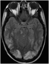
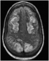
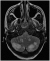
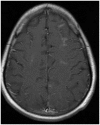
Results: Eight out of the 20 patients included in the study were on cyclosporine or tacrolimus based immunosuppressant therapy for kidney transplant. Four patients had severe hypertension at presentation. The most common MRI finding was high T2-fluid-attenuated inversion recovery (FLAIR) signal in the cortex and subcortical white matter of both cerebral hemispheres, particularly in the parietal and occipital lobes (n=16). The second most common MRI finding was increased leptomeningeal FLAIR signal (n=7). Out of seven patients with leptomeningeal signal, five demonstrated leptomeningeal enhancement as well. Four out of these seven patients had no other parenchymal findings.
Conclusion: Childhood PRES is commonly seen in the setting of immunosuppressant therapy for kidney transplant, severe hypertension and cancer treatment. There was high incidence of increased leptomeningeal FLAIR signal and leptomeningeal enhancement in our study. It supports the current theory of endothelial injury with increased microvascular permeability as the potential pathophysiology of PRES. Also, absence of elevated blood pressure in majority of the patients in our study supports the theory of direct endothelial injury by some agents leading to vasogenic edema.
Keywords: Magnetic resonance imaging; childhood; encephalopathy; posterior reversible encephalopathy syndrome.
© The Author(s) 2015.
Figures

References
-
- Sanders TG, Clayman DA, Sanchez-Ramos L, et al. Brain in eclampsia: MR imaging with clinical correlation. Radiology 1991; 180: 475–478. - PubMed
-
- Schwartz RB, Jones KM, Kalina P, et al. Hypertensive encephalopathy: findings on CT, MR imaging, and SPECT imaging in 14 cases. Am J Roentgenol 1992; 159: 379–383. - PubMed
MeSH terms
Substances
LinkOut - more resources
Full Text Sources
Other Literature Sources
Medical

