. 2015 Nov 3;66(18):1990-1999.
doi: 10.1016/j.jacc.2015.08.879.

Synergistic Effects of Combined Cell Therapy for Chronic Ischemic Cardiomyopathy

Affiliations

Synergistic Effects of Combined Cell Therapy for Chronic Ischemic Cardiomyopathy

Vasileios Karantalis et al. J Am Coll Cardiol. .

Abstract

Background: Both bone marrow-derived mesenchymal stem cells (MSCs) and c-kit(+) cardiac stem cells (CSCs) improve left ventricular remodeling in porcine models and clinical trials. Using xenogeneic (human) cells in immunosuppressed animals with acute ischemic heart disease, we previously showed that these 2 cell types act synergistically.

Objectives: To more accurately model clinical applications for heart failure, this study tested whether the combination of autologous MSCs and CSCs produce greater improvement in cardiac performance than MSCs alone in a nonimmunosuppressed porcine model of chronic ischemic cardiomyopathy.

Methods: Three months after ischemia/reperfusion injury, Göttingen swine received transendocardial injections with MSCs alone (n = 6) or in combination with cardiac-derived CSCs (n = 8), or placebo (vehicle; n = 6). Cardiac functional and anatomic parameters were assessed using cardiac magnetic resonance at baseline and before and after therapy.

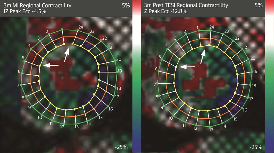
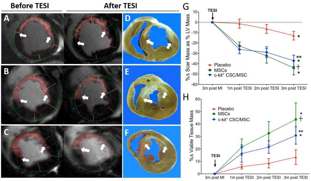
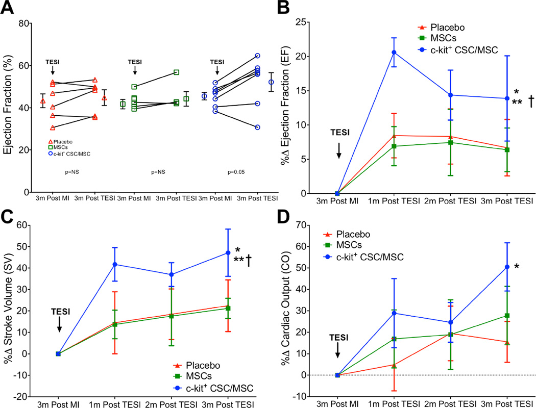
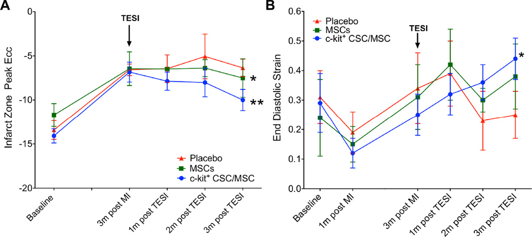
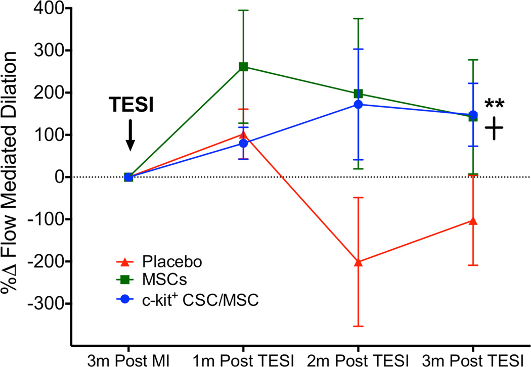
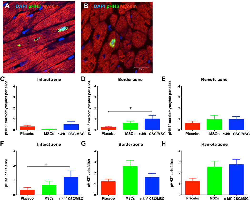
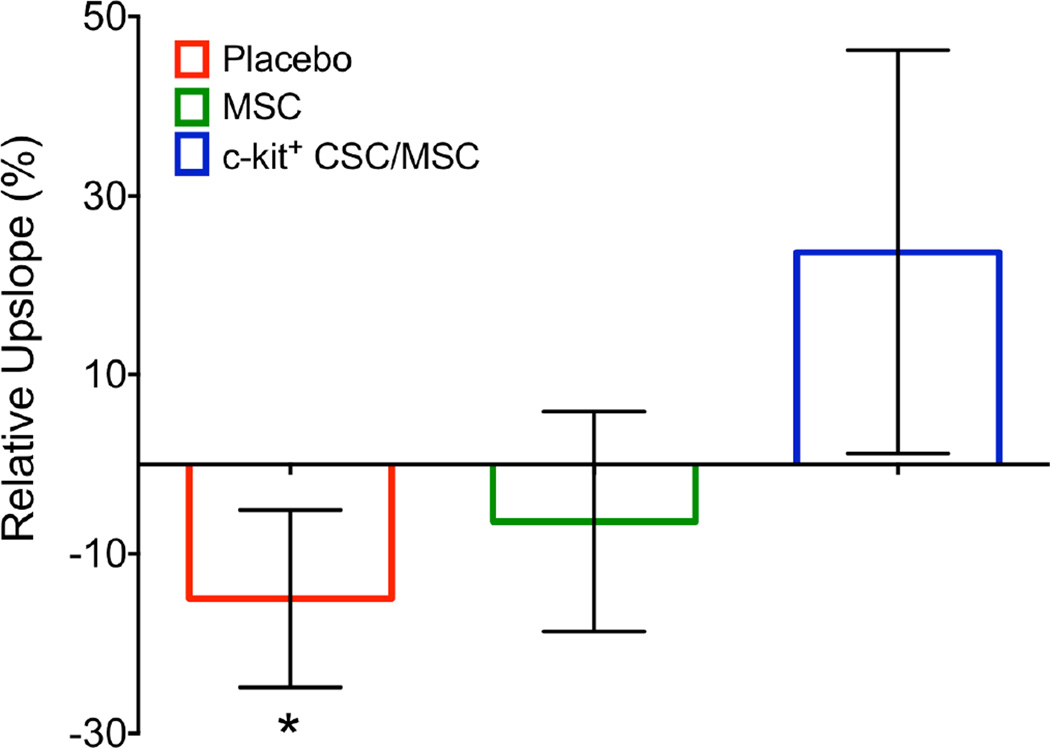
Results: Both groups of cell-treated animals exhibited significantly reduced scar size (MSCs -44.1 ± 6.8%; CSC/MSC -37.2 ± 5.4%; placebo -12.9 ± 4.2%; p < 0.0001), increased viable tissue, and improved wall motion relative to placebo 3 months post-injection. Ejection fraction (EF) improved (MSCs 2.9 ± 1.6 EF units; CSC/MSC 6.9 ± 2.8 EF units; placebo 2.5 ± 1.6 EF units; p = 0.0009), as did stroke volume, cardiac output, and diastolic strain only in the combination-treated animals, which also exhibited increased cardiomyocyte mitotic activity.

Conclusions: These findings illustrate that interactions between MSCs and CSCs enhance cardiac performance more than MSCs alone, establish the safety of autologous cell combination strategies, and support the development of second-generation cell therapeutic products.

Keywords: cardiac; combination therapy; heart failure; mesenchymal stem cell.


Conflict of interest statement

The other authors report no conflicts

Figures

FIGURE 1
FIGURE 1. Antifibrotic Effects Post-TESI
Short-axis sections of delayed enhancement cardiac magnetic resonance (A–C) depict the infarct extension (scar = red with white arrows) before treatment and, as seen in comparable gross pathology sections (D–F) 3 months following transendocardial stem cell injection (TESI). While TESI with placebo (n = 6) increased scar size from 7.2 g to 9.0 g (A,D), scar reductions occurred with autologous MSC (n = 5) from 9.7 g to 5.9 g (B,E) and autologous combination of ckit+ CSC/ MSC (n = 7) from 8.9 g to 5.8 g (C,F). (G) Cell-treated groups have similar scar size reduction (between-group comparison 2-way analysis of variance [ANOVA] p < 0.0001) and (H) increased viable tissue (between-group comparison 2-way ANOVA p = 0.0002). Graphs = mean ± SEM. *p < 0.05 within-group repeated measures 1-way ANOVA; 2-way ANOVA between-group comparison and Tukey's multicomparison test **p < 0.05 CSC/MSC vs. placebo at 1, 2, and 3 months post-TESI and +p < 0.05 MSC vs. placebo at 1, 2, and 3 months post-TESI. CSC = cardiac stem cell; LV = left ventricular; MSC = mesenchymal stem cell; MI = myocardial infarction.
FIGURE 2
FIGURE 2. EF Improvement Post-TESI
Change in ejection fraction (EF) for individual animals for EF units (A) and as a percent change (B; p = 0.01) post-TESI. Accompanying this EF restoration was a substantial improvement in the CSC/MSC group in (C) stroke volume (p = 0.008) and (D) cardiac output, which increased only in the CSC/MSC group (p = 0.007). Graphs represent mean ± SEM. *p < 0.05 1-way ANOVA, 3 months post-MI vs. 1, 2, and 3 months post TESI with CSC/MSC; **p < 0.05 CSC/MSC vs. placebo; †p < 0.05 CSC/MSC vs. MSCs. CO = cardiac output; other abbreviations as in Figure 1.
FIGURE 3
FIGURE 3. Contractility and Diastolic Strain
(A) Circumferential strain rate (peak Ecc) in the infarct zone improved in both cell-treated animal groups, but not the placebo group (*MSC: p = 0.04; ** CSC/MSC: p <0.004; between-group comparison: p = 0.1. (B) Diastolic strain increased only in the combination-treated animals (*p = 0.04), remaining unchanged in the MSC (p = NS) and placebo (p = NS) groups (between-group comparison: p = 0.9). Ecc = Eulerian circumferential shortening strain; other abbreviations as in Figure 1.
FIGURE 4
FIGURE 4. Endothelial Function
Flow-mediated dilation improved endothelial function in both cell-treated groups CSC/MSC vs. MSCs: **p <0.05; MSCs vs. placebo: +p < 0.05. Abbreviations as in Figure 1.
FIGURE 5
FIGURE 5. Treatment-enhanced Myocardial Mitotic Activity
Confocal microscopy depicts increased mitotic activity of endogenous cardiomyocytes (pHH3+ nuclei) in border (A) and remote (B) zones in cell-treated hearts at 3 months post-TESI. Based on the average number of pHH3+ mitotic cells within the myocardium per slide per group in the infarct (C), border (D), and remote (E) zones, combination cell therapy significantly increased myocardial mitotic activity in the border zone compared to placebo (*p = 0.05). (F, G, H) According to the average number of pHH3+ cardiomyocytes per slide per group in the infarct (F), border (G), and remote (H) zones, combination therapy produced significant increases in cardiomyocytes in the infarct zone (TESI site) compared to placebo (*p = 0.05). DAPI = 4',6-diamidino-2-phenylindole; PHH3 = phosphohistone H3; other abbreviations as in Figure 1.
FIGURE 6
FIGURE 6. Infarct Zone Perfusion
Using contrast-enhanced cardiac magnetic resonance images there was a trend towards deterioration of tissue perfusion in the infarct zone with placebo (−15.0 ± 9.9%;*p = 0.07). This decline did not occur in the cell treatment groups (MSC −6.4 ± 12.2%, p = 0.32; c kit+ CSC/MSC 23.7 ± 22.5%, p = 0.72). Bar graphs depict change in tissue perfusion from 3 months post-MI and 3 months post-TESI as measured by upslope analyses. Abbreviations as in Figure 1.
CENTRAL ILLUSTRATION
CENTRAL ILLUSTRATION. Combination Stem Cell Therapy for Heart Failure
Tagged harmonic phase cardiac magnetic resonance strain maps show significantly depressed regional function by peak Eulerian circumferential shortening strain (Ecc) at 3 months post-myocardial infarction (A) (white arrows). Red/white indicates weak contractility (more positive Ecc) and green/blue indicates vigorous contractility (more negative Ecc) in harmonic phase strain maps. At 3 months post-cell injection (B), infarct zone (IZ) contractility has improved (less red/white, more green/blue). TESI = transendocardial stem cell injection.

Comment in

References

    1. Hare JM, Fishman JE, Gerstenblith G, et al. Comparison of allogeneic vs autologous bone marrow-derived mesenchymal stem cells delivered by transendocardial injection in patients with ischemic cardiomyopathy: the POSEIDON randomized trial. JAMA. 2012;308:2369–2379. - PMC - PubMed
    1. Heldman AW, DiFede D, Fishman JE, et al. Transendocardial Mesenchymal Stem Cells and Mononuclear Bone Marrow Cells for Ischemic Cardiomyopathy: The TAC-HFT Randomized Trial. JAMA. 2014;311:62–73. - PMC - PubMed
    1. Karantalis V, Hare JM. Use of mesenchymal stem cells for therapy of cardiac disease. Circ Res. 2015;116:1413–1430. - PMC - PubMed
    1. Williams AR, Hare JM. Mesenchymal stem cells: biology, pathophysiology, translational findings, and therapeutic implications for cardiac disease. Circ Res. 2011;109:923–940. - PMC - PubMed
    1. Amado LC, Saliaris AP, Schuleri KH, et al. Cardiac repair with intramyocardial injection of allogeneic mesenchymal stem cells after myocardial infarction. Proc Natl Acad Sci U S A. 2005;102:11474–11479. - PMC - PubMed

Publication types

MeSH terms