Review
. 2015 Dec;34(4):643-90.
doi: 10.1007/s10555-015-9588-7.

Tumor cell survival pathways activated by photodynamic therapy: a molecular basis for pharmacological inhibition strategies

Affiliations
Review

Tumor cell survival pathways activated by photodynamic therapy: a molecular basis for pharmacological inhibition strategies

Mans Broekgaarden et al. Cancer Metastasis Rev. 2015 Dec.

Abstract

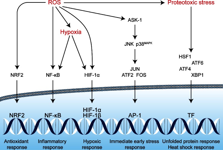
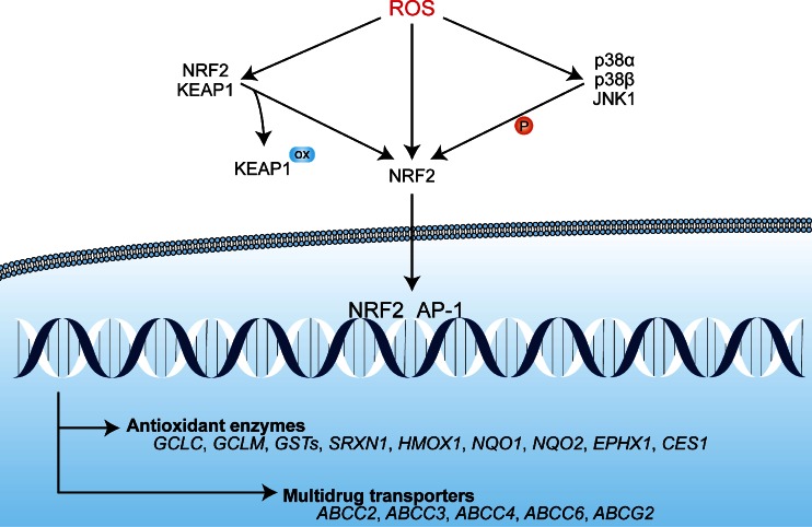
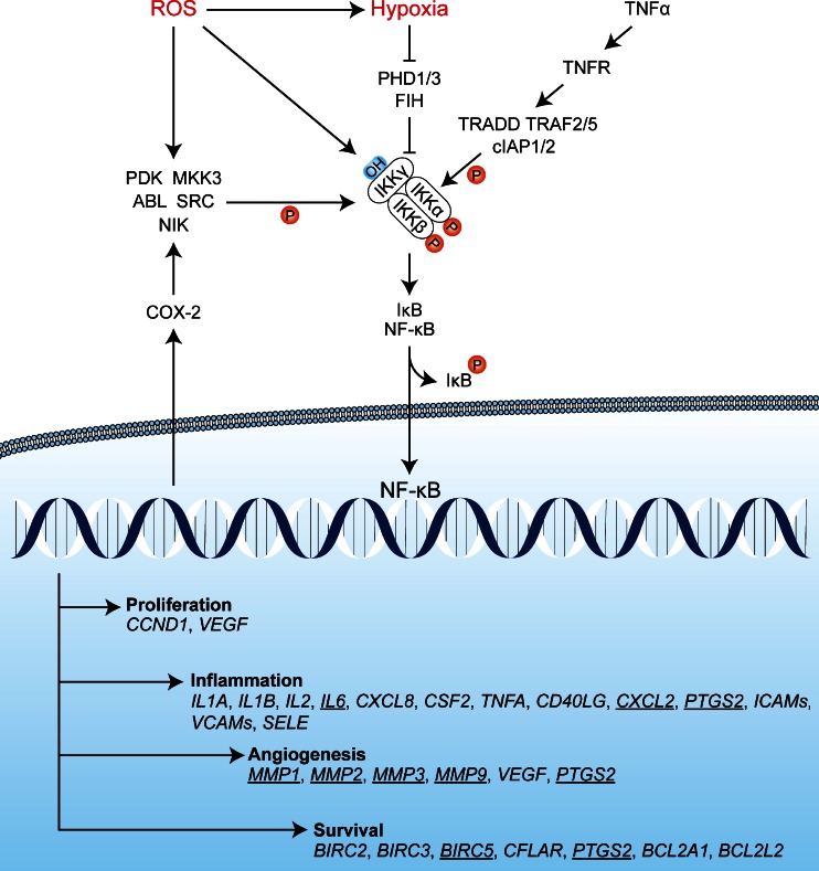
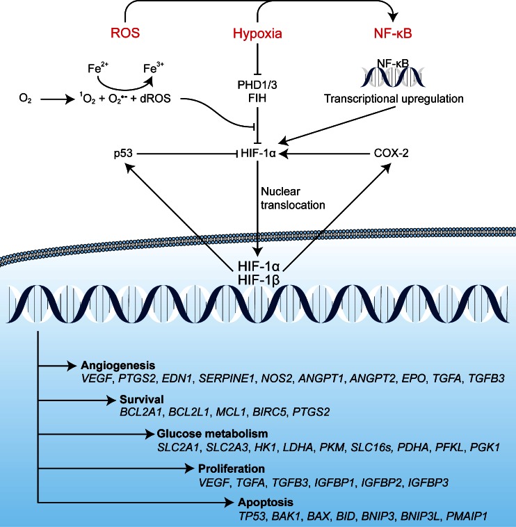
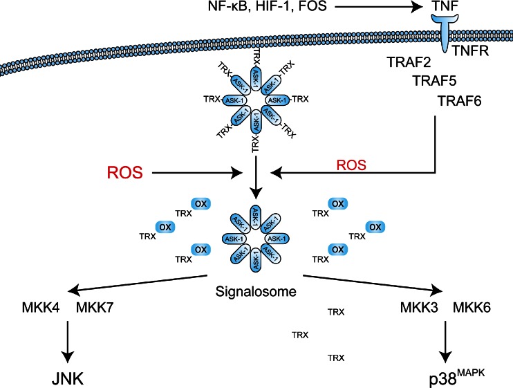
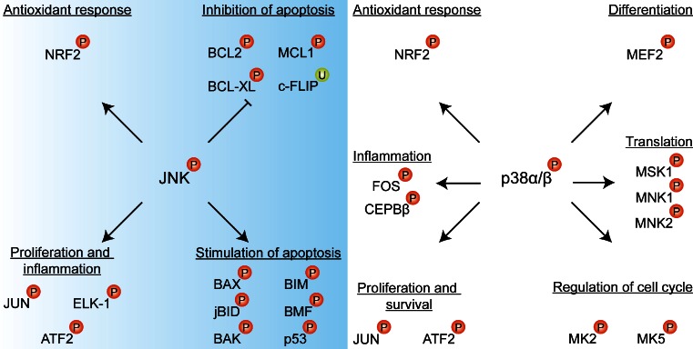
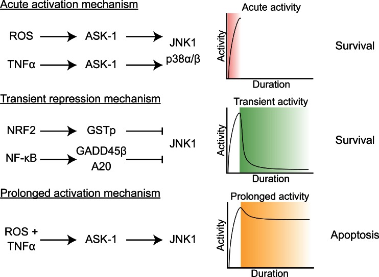
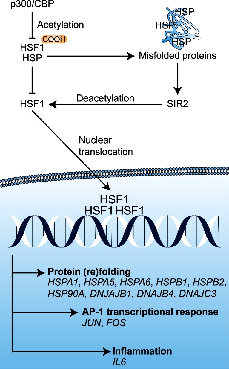
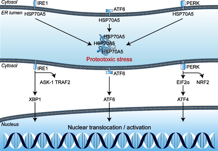
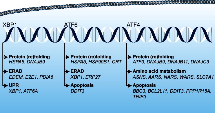
Photodynamic therapy (PDT) has emerged as a promising alternative to conventional cancer therapies such as surgery, chemotherapy, and radiotherapy. PDT comprises the administration of a photosensitizer, its accumulation in tumor tissue, and subsequent irradiation of the photosensitizer-loaded tumor, leading to the localized photoproduction of reactive oxygen species (ROS). The resulting oxidative damage ultimately culminates in tumor cell death, vascular shutdown, induction of an antitumor immune response, and the consequent destruction of the tumor. However, the ROS produced by PDT also triggers a stress response that, as part of a cell survival mechanism, helps cancer cells to cope with the PDT-induced oxidative stress and cell damage. These survival pathways are mediated by the transcription factors activator protein 1 (AP-1), nuclear factor E2-related factor 2 (NRF2), hypoxia-inducible factor 1 (HIF-1), nuclear factor κB (NF-κB), and those that mediate the proteotoxic stress response. The survival pathways are believed to render some types of cancer recalcitrant to PDT and alter the tumor microenvironment in favor of tumor survival. In this review, the molecular mechanisms are elucidated that occur post-PDT to mediate cancer cell survival, on the basis of which pharmacological interventions are proposed. Specifically, pharmaceutical inhibitors of the molecular regulators of each survival pathway are addressed. The ultimate aim is to facilitate the development of adjuvant intervention strategies to improve PDT efficacy in recalcitrant solid tumors.

Keywords: Antioxidant response; Apoptosis signaling kinase 1; ER stress; Heat shock factor 1; Inflammatory response; Proteotoxic stress.


Figures

Fig. 1
Fig. 1
a Overview of clinically obtained complete response rates with PDT of actinic keratoses (AK), skin cancers (SC), early stage central lung cancers (ECLC), esophageal malignancies (EM), nasopharyngeal carcinoma (NPC), and bladder cancer (BC). SC included (nodular) basal cell carcinomas and squamous cell carcinomas [9]. EM included Barrett’s esophagus, low-grade dysplasia, high-grade dysplasia, and esophageal cancer [10]. BC included carcinoma in situ, recurrent superficial bladder cancer, and early stage lesions [11]. Complete response rates were averaged using the longest time interval in each study. b Average of the median survival time postdiagnosis of extrahepatic cholangiocarcinoma patients treated with PDT or left untreated (control) [12]. Adjuvant treatments, type of photosensitizer, light source, and light dose were not taken into account, as a result of which no statistical analyses were performed
Fig. 2
Fig. 2
Reactive oxygen species (ROS)-induced activation of cell survival-related signal transduction pathways in cancer cells following photodynamic therapy (PDT). PDT induces vascular shutdown and oxidation of proteins, which results in hypoxia and proteotoxic stress, respectively. ROS directly trigger the NRF2-mediated antioxidant response and the ASK1-induced immediate early stress response. Hypoxia and ROS are both involved in the activation of the NF-κB inflammatory response and the HIF-1 hypoxic response. The proteotoxic stress response is characterized by the activation of several transcription factors (TF), including HSF1, ATF4, ATF6, and XBP1
Fig. 3
Fig. 3
The activation mechanism of NRF2 and downstream transcription events. Under normophysiological conditions, NRF2 is sequestered in an inactive cytoplasmic complex with KEAP1. Under oxidative stress conditions, ROS mediate the oxidation (ox) of essential cysteines in the NRF2-binding domain of KEAP1, which deters complex formation. NRF2 can be additionally oxidized at Cys183 by ROS under prooxidative conditions, which enables its nuclear translocation. Moreover, ROS can activate the ASK1 pathway, in which the MAPKs JNK1 and p38α/β phosphorylate (P) NRF2 at Ser40, leading to its activation. Subsequently, NRF2 translocates to the nucleus where it dimerizes with AP-1 transcription factors (Section 3.4.2) and initiates the transcription of antioxidant enzymes (e.g., glutathione synthesis) and multidrug transporters (ABCC2, ABCC3, ABCC4, ABCC6 and ABCG2)
Fig. 4
Fig. 4
Potential activation mechanisms of NF-κB in response to PDT. ROS may activate IKK directly by oxidizing redox-sensitive cysteines on IKKγ. Alternatively, IKKα/β may be phosphorylated by kinases such as PKD, ABL, SRC, NIK, and/or MKK3 in response to oxidative stress. Hypoxia is likely a coactivator of the IKK complex, since depletion of oxygen (O2 → ROS) renders PHD1/3 and FIH dysfunctional, as a result of which hydroxylation of IKKγ cannot occur and IKKγ is no longer targeted for proteasomal degradation by VHL-mediated polyubiquitination. Finally, in the presence of TNF-α, the TNFR becomes activated and triggers the assembly of a complex in which TRADD, TRAF2/5, and cIAP1/2 promote the phosphorylation of IKKα/β. A stabilized and activated IKK complex phosphorylates IκB, which dissociates from the NF-κB complex and relieves its sequestration in the cytoplasm. Upon release, NF-κB translocates to the nucleus to induce a transcriptional response that promotes proliferation, inflammation, angiogenesis, and survival. Via COX-2, EGFR signaling activates of a variety of kinases (e.g., PKD, MKK3, ABL, SRC, NIK) that in turn phosphorylate and activate the IKK complex. Active NF-κB transcription factors induce the transcription of genes involved in proliferation, inflammation, angiogenesis, and survival. The underlined genes have been extensively investigated in relation to PDT and are discussed in detail in the main text. CCND1 encodes cyclin D1, PTGS2 encodes COX-2, and BIRC5 encodes survivin
Fig. 5
Fig. 5
Activation of HIF-1α after PDT is mediated by several pathways. PDT-induced hypoxia due to immediate O2 depletion as well as vascular shutdown prevents HIF-1α hydroxylation by PHDs and FIH, which is an O2-dependent process. Furthermore, ROS-mediated oxidation of Fe2+ in the catalytic center of PHDs and FIH disables the enzymatic activity of these proteins. Both events lead to HIF-1α stabilization, translocation from the cytosol to the nucleus, complexation with HIF-1β, and transcriptional upregulation of numerous target genes containing an HRE in the promoter region. HIF-1α transcription is also upregulated by PDT-activated NF-κB, which increases HIF-1α protein levels. A negative feedback loop for HIF-1α exists via the upregulation of p53 by activated HIF-1, which targets HIF-1α for proteasomal degradation in the presence of DNA damage. A positive feedback loop exists via the upregulation of COX-2 by activated HIF-1 and NF-κB. COX-2 is involved in the production of PGE2 that plays a role in the transactivation of HIF-1α. After nuclear translocation, dimerization, and DNA binding to HRE sequences, HIF-1 transcription factors facilitate the upregulation of a plethora of genes involved in angiogenesis, survival, glucose metabolism, proliferation, and apoptosis. Other pathways are affected as well, but only those most relevant for PDT are depicted
Fig. 6
Fig. 6
Activation mechanisms of the ASK1 signaling pathway leading to JNK and p38MAPK phosphorylation. ROS can directly or indirectly (via GSH) oxidize the TRX subunits (TRX-ox) of the inactivated signalosome complex. Upon oxidation and subsequent dissociation of TRX, the ASK1 heteromer autophosphorylates and initiates downstream signaling. MKK4/7 are kinases responsible for the activation of JNK proteins, whereas MKK3/6 phosphorylate and activate the p38MAPKs. Downstream of the NF-κB, HIF-1, and FOS pathways, tumor cells and immune cells produce TNF-α. TNF-α binds the TNFR, which activates intracellular TRAF. These TRAFs stimulate the production of mitochondrial ROS and NADPH oxidase 1-derived ROS that stimulate the dissociation of TRX from the signalosome, but also bind TRX to prevent reassociation of TRX with ASK1
Fig. 7
Fig. 7
JNK1 exerts kinase activity on several transcription factors and BCL2 family proteins through phosphorylation (P) or ubiquitination (U). Proliferation and inflammation are induced by phosphorylating members of the JUN and ATF2 protein family, as well as ELK-1. The NRF2 antioxidant response is triggered via phosphorylation of NRF2 at Ser40. Apoptosis is stimulated via the phosphorylation of BAX, BAK, BIM, BID, and BMF proteins as well as via p53 activation. Antiapoptotic proteins are also phosphorylated (and inactivated) by JNK1, which include BCL2, BCL-XL, and MCL-1, or ubiquitinated (c-FLIP). The kinase functions of p38α/β entail a similar effect on the NRF2 antioxidant response pathway as reported for JNK1. Inflammation is stimulated via phosphorylation of the AP-1 transcription factors of the FOS and C/EBP protein family. Proliferation and survival are promoted via activation of JUN and ATF2. Translation of newly transcribed genes is facilitated by phosphorylation of MSK1, MNK1, and MNK2. Differentiation is mediated via MEF2 activation by p38α/β. Additionally, p38α/β regulate the cell cycle by phosphorylation of MK2 and MK5
Fig. 8
Fig. 8
The ambivalent effects of the ASK1 pathway are dictated by the cross-talk between various pathways and the prevailing biochemical conditions. The primary activation mechanism of the ASK1 pathway by PDT emanates from oxidative stress or TNF-α signaling, leading to the acute, survival-promoting activity of JNK1 and p38α/β. Subsequently, downstream of the NRF2 and the NF-κB pathways, negative regulators of JNK1 and p38α/β are produced/activated that modulate the transient activation pattern of these kinases and thus promote cell survival. Whenever ROS and TNF-α signaling occur simultaneously, or whenever these stress signals endure, prolonged JNK1 and p38α/β activation promotes apoptosis
Fig. 9
Fig. 9
Mechanism of HSF1 activation. Monomeric HSF1 is kept inactive in the cytosol via acetylation by p300/CREB binding protein. The cytosolic monomers interact with HSP40, 70, or 90, which prevents DNA binding and transactivation. Proteotoxic stress titrates HSPs away from HSF1 and stress-induced SIR2 deacetylates the HSF1 monomers. Subsequently, HSF1 forms homotrimers, translocates to the nucleus, and binds genomic heat shock elements to facilitate target gene expression. The target gene products are mainly involved in protein (re)folding, but also enforce the immediate early stress response and inflammation
Fig. 10
Fig. 10
Activation mechanism of the ER stress response as a result of proteotoxic stress. Unfolded protein detectors IRE1, PERK, and ATF6 are sequestered by HSP70A5. Upon proteotoxic stress due to protein oxidation, misfolding, or formation of protein aggregates, HSP70A5 is recruited toward the misfolded proteins. The ER stress detectors are subsequently activated, leading to the initiation of XBP1, ATF4, and ATF6 transcription factor function
Fig. 11
Fig. 11
Transcriptional regulation of genes induced by XBP1, ATF6, and ATF4 in response to proteotoxic stress. XBP1 stimulates protein (re)folding, ERAD, and amplifies the UPR. ATF6 also promotes protein (re)folding and ERAD, but also stimulates apoptosis by upregulating DDIT3 (CHOP). ATF4 ameliorates proteotoxic stress by upregulating ATF3 and a plethora of DNAJ genes (encoding various isoforms of HSP40). ATF4 additionally upregulates genes involved in amino acid metabolism that include, but are not limited to, asparagine synthetase (ASNS), alanyl-tRNA synthetase (AARS), asparagyl-tRNA synthetase (NARS), tryptophanyl-tRNA synthetase (WARS), and the cationic amino acid transporter SLC7A1. ATF4 additionally upregulates proapoptotic genes BBC3, BCL2L11, DDIT3, PPP1R15A, and TRIB3

References

    1. Triesscheijn M, Baas P, Schellens JHM, Stewart FA. Photodynamic therapy in oncology. The Oncologist. 2006;11:1034–1044. doi: 10.1634/theoncologist.11-9-1034. - DOI - PubMed
    1. Dolmans DEJGJ, Fukumura D, Jain RK. Photodynamic therapy for cancer. Nature Reviews Cancer. 2003;3:380–387. doi: 10.1038/nrc1071. - DOI - PubMed
    1. Grant WE, Speight PM, Hopper C, B SG. Photodynamic therapy: an effective, but non-selective treatment for superficial cancers of the oral cavity. International Journal of Cancer. 1997;71:937–942. doi: 10.1002/(SICI)1097-0215(19970611)71:6<937::AID-IJC4>3.0.CO;2-Z. - DOI - PubMed
    1. Kübler A, Niziol C, Sidhu M, Dünne A, Werner JA. Analysis of cost effectiveness of photodynamic therapy with Foscan (Foscan PDT) in comparison to palliative chemotherapy in patients with advanced head-and-neck tumors in Germany. Laryngo- Rhino- Otologie. 2005;84:725–732. doi: 10.1055/s-2005-861048. - DOI - PubMed
    1. Ris HB, Altermatt HJ, Inderbitzi R, Hess R, Nachbur B, Stewart JC, Wang Q, Lim CK, Bonnett R, B MC. Photodynamic therapy with chlorins for diffuse malignant mesothelioma: initial clinical results. British Journal of Cancer. 1991;48:1273–1283. - PMC - PubMed

MeSH terms