. 2015 Oct 26;125(12):4514-28.
doi: 10.1172/JCI82719.

Endothelial-to-mesenchymal transition drives atherosclerosis progression

Endothelial-to-mesenchymal transition drives atherosclerosis progression

Pei-Yu Chen et al. J Clin Invest. .

Abstract

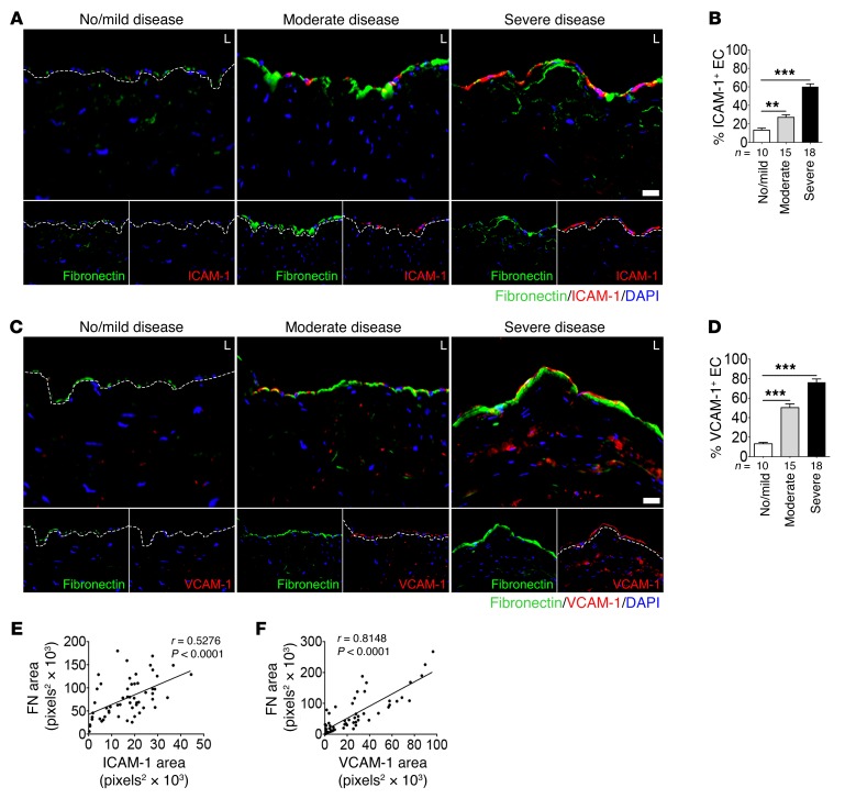
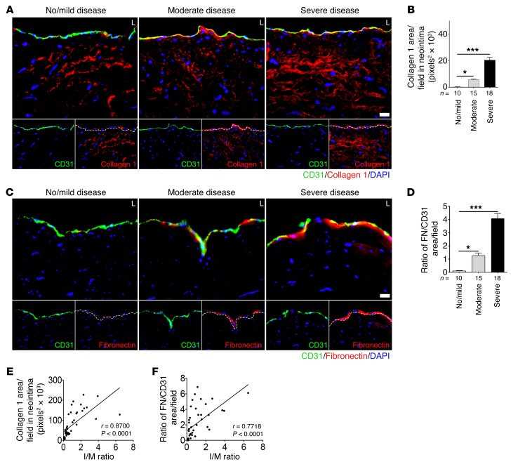
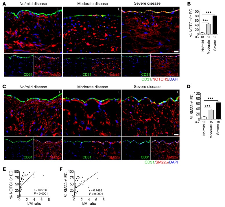
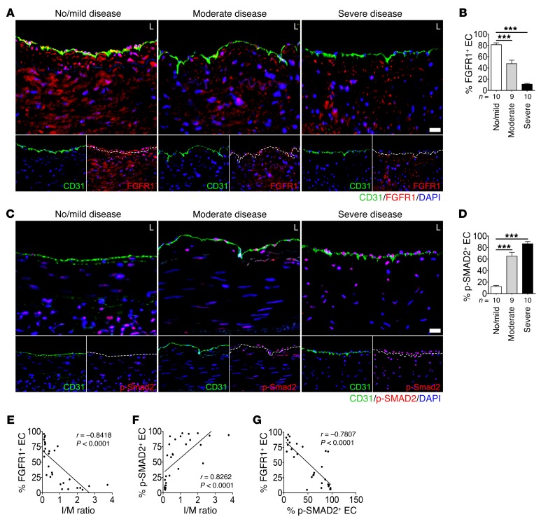
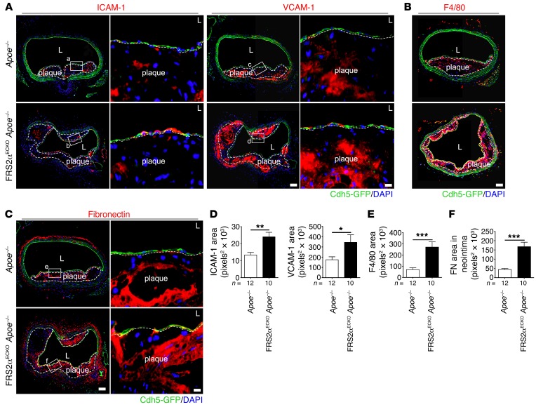
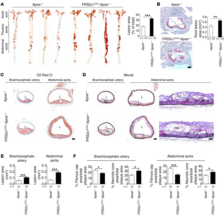
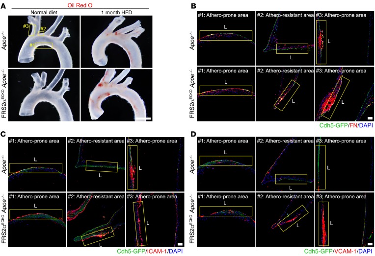
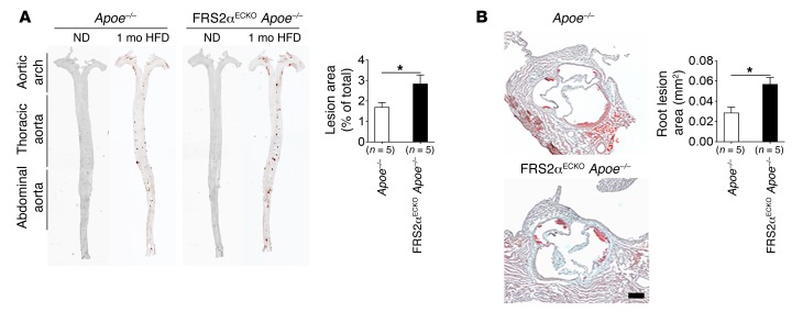
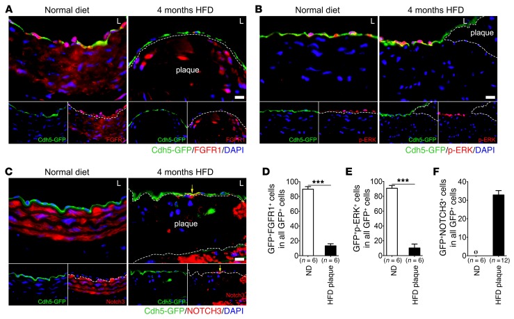
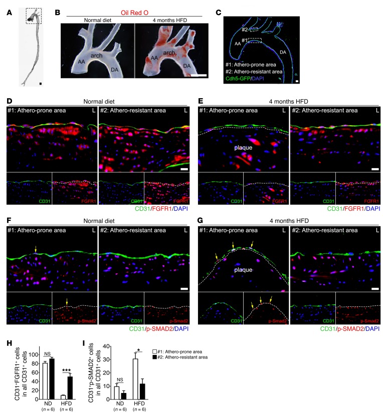
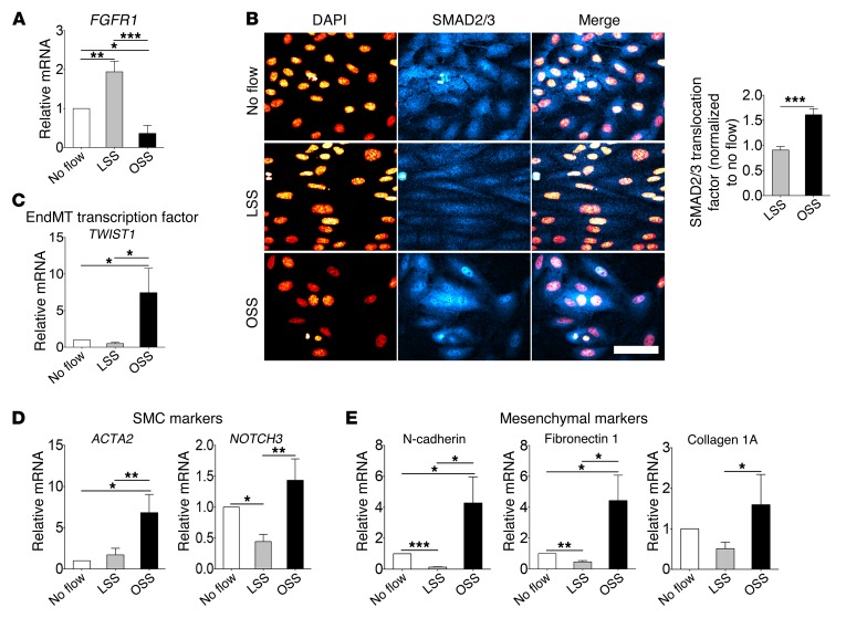
The molecular mechanisms responsible for the development and progression of atherosclerotic lesions have not been fully established. Here, we investigated the role played by endothelial-to-mesenchymal transition (EndMT) and its key regulator FGF receptor 1 (FGFR1) in atherosclerosis. In cultured human endothelial cells, both inflammatory cytokines and oscillatory shear stress reduced endothelial FGFR1 expression and activated TGF-β signaling. We further explored the link between disrupted FGF endothelial signaling and progression of atherosclerosis by introducing endothelial-specific deletion of FGF receptor substrate 2 α (Frs2a) in atherosclerotic (Apoe(-/-)) mice. When placed on a high-fat diet, these double-knockout mice developed atherosclerosis at a much earlier time point compared with that their Apoe(-/-) counterparts, eventually demonstrating an 84% increase in total plaque burden. Moreover, these animals exhibited extensive development of EndMT, deposition of fibronectin, and increased neointima formation. Additionally, we conducted a molecular and morphometric examination of left main coronary arteries from 43 patients with various levels of coronary disease to assess the clinical relevance of these findings. The extent of coronary atherosclerosis in this patient set strongly correlated with loss of endothelial FGFR1 expression, activation of endothelial TGF-β signaling, and the extent of EndMT. These data demonstrate a link between loss of protective endothelial FGFR signaling, development of EndMT, and progression of atherosclerosis.


Figures

Figure 11
Figure 11. Mesenchymal and inflammatory marker expression in the endothelium of human coronary arteries.
Left main coronary arteries from patients with no/mild (n = 10), moderate (n = 15), and severe (n = 18) CAD were evaluated. (A and C) Representative images of immunofluorescence staining of left main coronary arteries for fibronectin (green), ICAM-1 (red), and VCAM-1 (red) in patients. Nuclei were stained with DAPI (blue). Scale bar: 16 μm. (B) Percentage of ICAM-1+ ECs in the lumen (**P < 0.01 compared moderate disease to no/mild disease; ***P < 0.001 compared moderate disease to no/mild disease; ***P < 0.001 compared severe disease to no/mild disease). (D) Percentage of VCAM-1+ ECs in the lumen (***P < 0.001 compared moderate disease to no/mild disease; ***P < 0.001 compared severe disease to no/mild disease; 1-way ANOVA with Newman-Keuls post-hoc test for multiple comparison correction). (E and F) Scatter plots of fibronectin area and ICAM-1 and VCAM-1 area per field in the lumen. The corresponding Spearman’s correlation coefficient (r) between fibronectin area and ICAM-1 and VCAM-1 area per field and the P value are shown.
Figure 10
Figure 10. Mesenchymal marker expression in the endothelium of human coronary arteries.
Left main coronary arteries from patients with no/mild (n = 10), moderate (n = 15), and severe (n = 18) CAD were evaluated. (A and C) Representative images of immunofluorescence staining of left main coronary arteries for CD31 (green), collagen 1 (red), and fibronectin (red) in patients. Nuclei were stained with DAPI (blue). Scale bar: 16 μm. (B) Measurement of the collagen 1 area in the neointima per field (*P < 0.05 compared moderate disease to no/mild disease; ***P < 0.001 compared severe disease to no/mild disease; 1-way ANOVA with Newman-Keuls post-hoc test for multiple comparison correction). (D) Ratio of fibronectin area to CD31 area per field in the lumen (*P < 0.05 compared moderate disease to no/mild disease; ***P < 0.001 compared severe disease to no/mild disease; 1-way ANOVA with Newman-Keuls post-hoc test for multiple comparison correction). (E and F) Scatter plots of collagen 1 area in the neointima per field and the ratio of fibronectin area to CD31 area per field in the lumen and the I/M ratio. The corresponding Spearman’s correlation coefficient (r) between collagen 1 area in the neointima per field and the ratio of fibronectin area to CD31 area per field and the I/M ratio and the P value are shown.
Figure 9
Figure 9. Smooth muscle marker expression in the endothelium of human coronary arteries.
Left main coronary arteries from patients with no/mild (n = 10), moderate (n = 15), and severe (n = 18) CAD were evaluated. (A and C) Representative images of immunofluorescence staining of left main coronary arteries for CD31 (green), NOTCH3 (red), and SM22α (red). Nuclei were stained with DAPI (blue). Scale bar: 16 μm. (B and D) Percentage of NOTCH3+ ECs or SM22α+ ECs in the lumen (***P < 0.001 compared with no/mild disease; 1-way ANOVA with Newman-Keuls post-hoc test for multiple comparison correction). (E and F) Scatter plots of NOTCH3+ ECs or SM22α+ ECs in the lumen and the I/M ratio. The corresponding Spearman’s correlation coefficient (r) between NOTCH3+ ECs or SM22α+ luminal ECs and the I/M ratio and the P value are shown.
Figure 8
Figure 8. FGFR1 expression and SMAD2 phosphorylation in ECs in human coronary arteries.
Immunocytochemical analysis of (A) FGFR1 and (C) p-SMAD2 expression in the endothelium of left main coronary arteries of patients with no/mild (n = 10), moderate (n = 9), and severe (n = 10) CAD. Representative images of immunofluorescence staining for CD31 (green) and FGFR1 (red) or p-SMAD2 (red). Nuclei were stained with DAPI (blue). Scale bar: 16 μm. (B and D) Percentage of FGFR1+ ECs or p-SMAD2+ ECs in the lumen (***P < 0.001 compared with no/mild disease; 1-way ANOVA with Newman-Keuls post-hoc test for multiple comparison correction). (E and F) Scatter plots of FGFR1 or p-SMAD2 and the I/M ratio. The corresponding Spearman’s correlation coefficient (r) between FGFR1 or p-SMAD2 and the I/M ratio and the P value are shown. (G) Scatterplot of FGFR1 and p-SMAD2. The corresponding Spearman’s correlation coefficient (r) between FGFR1 and p-SMAD2 and the P value are shown.
Figure 7
Figure 7. Effect of endothelial FGF signaling suppression on inflammation and EndMT marker gene expression in mice.
(AC) Histological analysis of atherosclerotic plaques with anti–ICAM-1, anti–VCAM-1, anti-F4/80, and anti-fibronectin antibodies (Apoe–/– mice, n = 12; FRS2αECKO Apoe–/– mice, n = 10). Note circumferential plaque in FRS2αECKO Apoe–/– mice. Nuclei were counterstained with DAPI (blue). Scale bar: 62 μm (low-magnification images); 10 μm (high-magnification images). High-magnification images of the boxed areas are shown to the right. (DF) Measurement of F4/80, ICAM-1, VCAM-1, and fibronectin area (*P < 0.05; **P < 0.01; ***P < 0.001 compared with Apoe–/–; unpaired 2-tailed Student’s t test).
Figure 6
Figure 6. Effect of endothelial FGF signaling suppression on atherosclerosis in mice after 4 months of HFD.
(A) Microphotographs of aortas from Apoe–/– and FRS2αECKO Apoe–/– mice (24 weeks of age) after 4 months of HFD in the en face preparation after staining with Oil Red O and quantification of lesion areas (10 mice per group). All data are shown as mean ± SD (***P < 0.001 compared with Apoe–/–; unpaired 2-tailed Student’s t test). (B) Representative examples of cross sections from the aortic root stained with Oil Red O and quantification of aortic root lesion areas (12 mice per group). Scale bar: 200 μm. Mean ± SD (**P < 0.01 compared with Apoe–/–; unpaired 2-tailed Student’s t test). (C and D) Representative images of Oil Red O– or Movat-stained brachiocephalic arteries and abdominal aortas from Apoe–/– and FRS2αECKO Apoe–/– mice (Apoe–/– mice, n = 12; FRS2αECKO Apoe–/– mice, n = 10). Scale bar: 100 μm. High-magnification images of the lesion shown in boxed areas are shown to the right. Scale bar: 50 μm. NC, necrotic core. Note circumferential plaque in FRS2αECKO Apoe–/– mice. (E) Measurement of lesion area (***P < 0.001 compared with Apoe–/–; unpaired 2-tailed Student’s t test). (F) Quantifications of the extent of fibrous cap and necrotic areas in brachiocephalic arteries and abdominal aortas of Apoe–/– and FRS2αECKO Apoe–/– mice (*P < 0.05; ***P < 0.001 compared with Apoe–/–; unpaired 2-tailed Student’s t test).
Figure 5
Figure 5. Early onset of atherogenesis in FRS2αECKO Apoe–/– mice.
Apoe–/– and FRS2αECKO Apoe–/– mice were placed on a HFD and examined 4 weeks later. (A) Representative photomicrographs of Oil Red O–stained atherosclerotic lesions in the aortic arch before and after 4 weeks of HFD (5 mice per group). #1 and #3 denote atherosclerosis-prone areas, while #2 denotes an atherosclerosis-resistant area. Scale bar: 4 mm. (BD) Fibronectin (FN), ICAM-1, and VCAM-1 longitudinal sections of areas corresponding to boxes #1–#3 from A in Apoe–/– and FRS2αECKO Apoe–/– mice (5 mice per group). Nuclei were stained with DAPI (blue). Scale bar: 16 μm.
Figure 4
Figure 4. Effect of endothelial FGF signaling suppression on atherosclerosis in mice after 4 weeks of HFD.
Apoe–/– and FRS2αECKO Apoe–/– mice were placed on a HFD and examined 4 weeks later. (A) Microphotographs of aortas from Apoe–/– and FRS2αECKO Apoe–/– mice at 12 weeks of age in the en face preparation after staining with Oil Red O (5 mice per group) and quantification of lesion area. All data are shown as mean ± SD (*P < 0.05 compared with Apoe–/–; unpaired 2-tailed Student’s t test). (B) Representative examples of cross sections from the Oil Red O–stained aortic root (5 mice per group) and quantification of aortic root lesion areas. Scale bar: 200 μm. Mean ± SD. (*P < 0.05 compared with Apoe–/–; unpaired 2-tailed Student’s t test).
Figure 3
Figure 3. FGF signaling activity and EndMT extent in a mouse atherosclerosis model.
(A) Immunofluorescence staining of FGFR1 in aortas of Cdh5-CreERT2 mT/mG Apoe–/– mice (6 mice per group). Scale bar: 10 μm. (B) Cdh5-CreERT2 mT/mG Apoe–/– mice after 16 weeks on HFD (n = 6) or normal diet (n = 6) were injected i.v. with FGF2 (1 μg per mouse), and p-ERK activation was determined by immunofluorescence staining. Scale bar: 10 μm. (C) Immunofluorescence staining of NOTCH3 in aorta and plaque sections in Cdh5-CreERT2 mT/mG Apoe–/– mice (normal diet, n = 6; HFD, n = 12). Scale bar: 10 μm. (DF) Quantification of the number of luminal ECs expressing FGFR1, p-ERK, and NOTCH3 (***P < 0.001 compared with normal diet; unpaired 2-tailed Student’s t test). Yellow arrows indicate ECs expressing NOTCH3. Ø, not detected.
Figure 2
Figure 2. Shear stress downregulates FGFR1 expression and upregulates TGF-β signaling in vivo.
(A) Whole mouse aorta. Scale bar: 1 mm. (B) Representative photomicrographs of Oil Red O–stained aortic arch sections of Apoe–/– mice after 16 weeks on the normal diet (n = 3) or HFD (n = 3). Scale bar: 4 mm. (C) A high-magnification image of A, showing the aortic arch in the longitudinal section. AA, ascending aorta; DA, descending aorta. Scale bar: 160 μm. (DG) Immunofluorescence staining of CD31 (green), FGFR1 (red), and p-SMAD2 (red) in the aortic arches of Apoe–/– mice after 16 weeks on normal diet (n = 6) or HFD (n = 6). Atherosclerosis-prone areas are denoted by #1, while an atherosclerosis-resistant areas are denoted by #2. Yellow arrows indicate ECs expressing p-SMAD2. Nuclei were stained with DAPI (blue). L, lumen. Scale bar: 10 μm. (H and I) Quantification of the number of luminal EC expressing FGFR1 and p-SMAD2 (NS, not significant compared with #1; *P < 0.05 compared with #1; ***P < 0.001 compared with #1; unpaired 2-tailed Student’s t test).
Figure 1
Figure 1. Shear stress downregulates FGFR1 expression and upregulates TGF-β signaling in vitro.
(A and B) HUVECs were subject to 12 dynes/cm2 LSS or 1 ± 4 dynes/cm2 OSS for 16 hours. FGFR1 expression and SMAD2/3 nuclear translocation were examined by (A) qRT-PCR and (B) immunofluorescence staining. The bar graph of qRT-PCR results is representative of 4 independent experiments (*P < 0.05; **P < 0.01; ***P < 0.001; 1-way ANOVA with Newman-Keuls post-hoc test for multiple comparison correction). SMAD2/3 nuclear translocation images are representative of 3 independent experiments (***P < 0.001; unpaired 2-tailed Student’s t test). Scale bar: 50 μm. (CE) HUVECs were subject to 12 dynes/cm2 LSS or 1 ± 4 dynes/cm2 OSS for 48 hours. EndMT marker gene expression was examined by qRT-PCR. Bar graphs of qRT-PCR results are representative of 4 independent experiments (*P < 0.05; **P < 0.01; 1-way ANOVA with Newman-Keuls post-hoc test for multiple comparison correction).

References

    1. Conway DE, Schwartz MA. Flow-dependent cellular mechanotransduction in atherosclerosis. J Cell Sci. 2013;126(pt 22):5101–5109. - PMC - PubMed
    1. Hahn C, Schwartz MA. Mechanotransduction in vascular physiology and atherogenesis. Nat Rev Mol Cell Biol. 2009;10(1):53–62. doi: 10.1038/nrm2596. - DOI - PMC - PubMed
    1. Libby P, Ridker PM, Hansson GK. Progress and challenges in translating the biology of atherosclerosis. Nature. 2011;473(7347):317–325. doi: 10.1038/nature10146. - DOI - PubMed
    1. Libby P, Lichtman AH, Hansson GK. Immune effector mechanisms implicated in atherosclerosis: from mice to humans. Immunity. 2013;38(6):1092–1104. doi: 10.1016/j.immuni.2013.06.009. - DOI - PMC - PubMed
    1. Tabas I, Garcia-Cardena G, Owens GK. Recent insights into the cellular biology of atherosclerosis. J Cell Biol. 2015;209(1):13–22. doi: 10.1083/jcb.201412052. - DOI - PMC - PubMed

Publication types

MeSH terms