. 2015 Oct 26;125(12):4544-58.
doi: 10.1172/JCI82871.

Saturated phosphatidic acids mediate saturated fatty acid-induced vascular calcification and lipotoxicity

Saturated phosphatidic acids mediate saturated fatty acid-induced vascular calcification and lipotoxicity

Masashi Masuda et al. J Clin Invest. .

Abstract

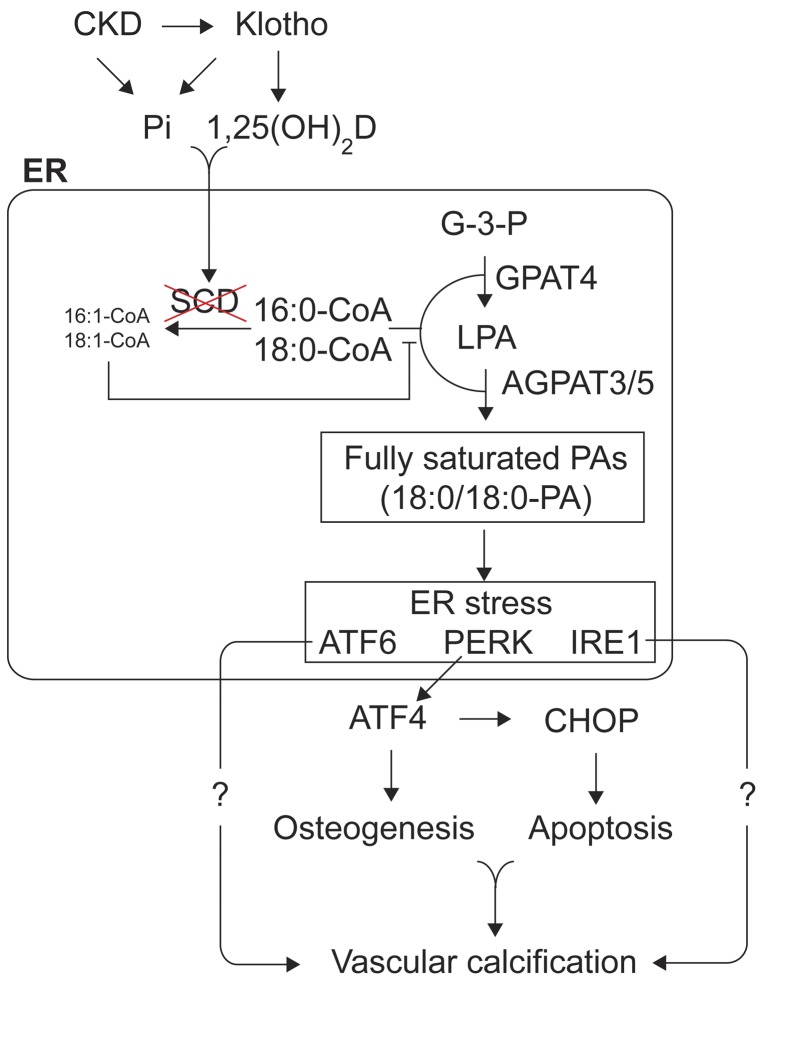
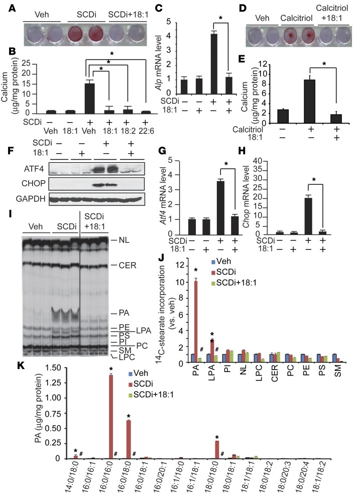
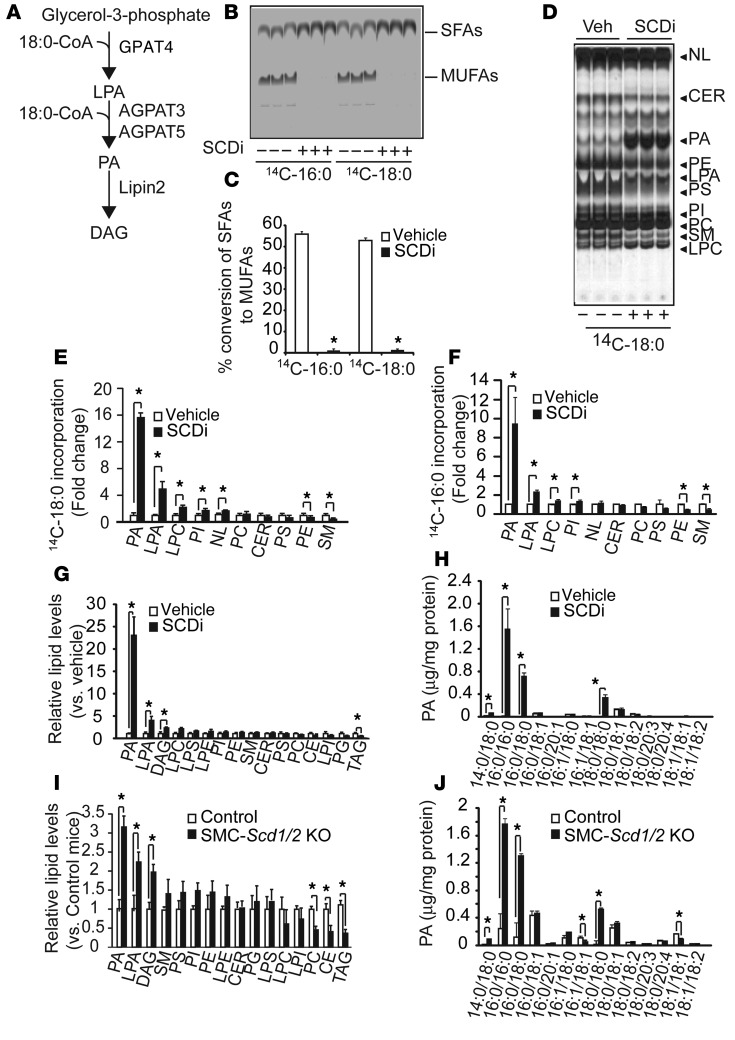
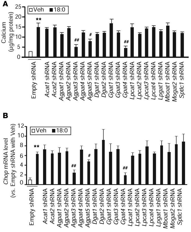
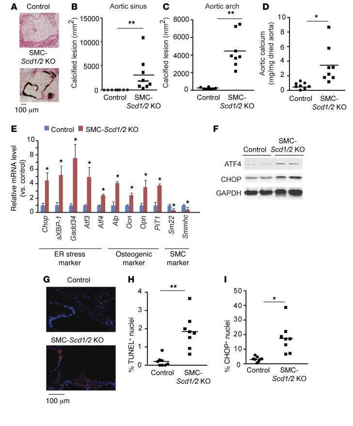
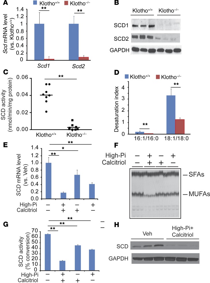
Recent evidence indicates that saturated fatty acid-induced (SFA-induced) lipotoxicity contributes to the pathogenesis of cardiovascular and metabolic diseases; however, the molecular mechanisms that underlie SFA-induced lipotoxicity remain unclear. Here, we have shown that repression of stearoyl-CoA desaturase (SCD) enzymes, which regulate the intracellular balance of SFAs and unsaturated FAs, and the subsequent accumulation of SFAs in vascular smooth muscle cells (VSMCs), are characteristic events in the development of vascular calcification. We evaluated whether SMC-specific inhibition of SCD and the resulting SFA accumulation plays a causative role in the pathogenesis of vascular calcification and generated mice with SMC-specific deletion of both Scd1 and Scd2. Mice lacking both SCD1 and SCD2 in SMCs displayed severe vascular calcification with increased ER stress. Moreover, we employed shRNA library screening and radiolabeling approaches, as well as in vitro and in vivo lipidomic analysis, and determined that fully saturated phosphatidic acids such as 1,2-distearoyl-PA (18:0/18:0-PA) mediate SFA-induced lipotoxicity and vascular calcification. Together, these results identify a key lipogenic pathway in SMCs that mediates vascular calcification.


Figures

Figure 8
Figure 8. Schematic diagram of SFA-dependent vascular calcification.
Increased SFAs through SCD inhibition leads to the accumulation of fully saturated PAs in the ER via de novo glycerolipid synthesis. The accumulation of fully saturated PAs induces ER stress responses such as ATF4-mediated osteogenesis and CHOP-mediated apoptosis, leading to vascular calcification. UFAs, however, competitively inhibit the formation of fully saturated PAs on the reaction of GPAT4, AGPAT3, and AGPAT5. Pi; inorganic phosphate; 1,2(OH)2D, calcitriol; G-3-P, Glycerol-3-phosphate.
Figure 7
Figure 7. Fully unsaturated, but not partially saturated or fully saturated PAs, induce mineralization, osteogenic differentiation, and ER stress.
(A) Fully saturated PAs are disfavor substrates for LIPIN2. The lysate of HEK293 cells infected with adenovirus containing human LIPIN2 was used as a LIPIN2 recombinant protein. LIPIN2 activity was measured as the conversion of PA to DAG using LC-MS/MS. The fully saturated PAs 18:0/18:0-PA and 16:0/16:0-PA)but not the partially saturated PAs 18:0/18:1-PA and 16:0/18:1-PA or the fully unsaturated PAs 18:1/18:1-PA and 18:2/18:2-PA dose-dependently induced (BD) mineralization and (EG) osteogenic differentiation of VSMCs. VSMCs were treated with each PA for 7 days in the presence of 2.0 mM phosphate. (B) Seven days after the treatments, the cells treated with PAs (20 μM) were stained with Alizarin red to identify calcium deposits. (C and D) Calcium was extracted with 0.6 N hydrochloric acid and analyzed using a colorimetric assay. (EG) Ocn, Opn, and PiT1 were quantified by qPCR. (HJ) Fully saturated PAs such as 18:0/18:0-PA, but not fully unsaturated PAs such as 18:1/18:1-PA, induce ER stress. VSMCs were treated with 10 μM PAs for 6 and 24 hours. Total protein extracts were subjected to immunoblot analysis with ATF4- and CHOP-specific antibodies. Atf4 and Chop mRNA levels were quantified by qPCR. (K) 18:0/18:0-PA induces mineralization of VSMCs through the activation of the ATF4-CHOP axis of the ER stress response. Atf4 and Chop knockdown MOVAS cells were treated with 10 μM 18:0/18:0-PA for 7 days. n = 6; *P < 0.05 (one-way ANOVA).
Figure 6
Figure 6. UFA cotreatment normalizes mineralization, osteogenic differentiation, ER stress, and PA accumulation.
(AC) UFAs blocked SCDi-induced mineralization (A and B) and osteogenic differentiation (C) of VSMCs. Human VSMCs were treated with 200 μM UFAs in the presence of 300 nM SCDi for 7 days in the presence of 2.0 mM inorganic phosphate. (A) Seven days after the treatments, the cells were stained with Alizarin red to identify calcium deposits. (B) Calcium content was analyzed using a colorimetric assay. (C) Alp mRNA levels were analyzed by qPCR. (D and E) Calcitriol-induced mineralization is blocked by 18:1n-9 supplementation. Human VSMCs were treated with 100 nM calcitriol in the presence of 200 μM 18:1n-9 for 7 days. (FH) UFAs such as 18:1n-9 completely blocked ER stress induced by SCDi. VSMCs were treated with SCDi acid for 24 hours. Total protein extracts were subjected to immunoblot analysis with ATF4- and CHOP-specific antibodies. Atf4 and Chop mRNA levels were quantified by qPCR. (I) Autoradiography and (J) quantification of 14C-18:0 incorporation into the lipid fraction. Human VSMCs were treated with 200 μM 18:1n-9 in the presence of 300 nM SCDi and 14C-18:0 (1 μCi). The black line in I indicates that the image was derived from noncontiguous lanes of the same plate. Lipids were separated on a boric acid–coated TLC. (K) Absolute levels of PA species in VSMCs cotreated with SCDi and 18:1n-9. Human VSMCs were treated with 200 μM 18:1n-9 in the presence of 300 nM SCDi for 24 hours. Each PA content was analyzed with LC-MS/MS. NL, neutral lipids. n = 4, *P < 0.01 vs. vehicle (Veh) and #P < 0.01 vs. SCDi (n = 3–6, one-way ANOVA).
Figure 5
Figure 5. SFAs were preferentially incorporated into PA and accumulated as fully saturated PAs in VSMCs in vitro and in vivo.
(A) Schematic representation of GPAT4, AGPAT3, and AGPAT5, which are localized in the ER. (B) Autoradiography and (C) quantification of SCD activity in human VSMCs treated with 300 nM of the SCDi CAY10566 using 14C-18:0 and 14C-16:0 as substrates (n = 6). VSMCs were treated with 14C-18:0 and 14C-16:0 for 6 hours in the presence/absence of SCDi. SFAs and MUFAs isolated from total cell lysate were separated on a silver nitrate–coated TLC. (D) Autoradiography and quantification of (E) 14C-18:0 and (F) 16:0 incorporation into the lipid fraction in human VSMCs treated with SCDi. Human VSMCs were pretreated with SCDi for 2 hours and incubated with 14C-18:0 and 14C-16:0 for 6 hours in the presence/absence of SCDi. Total lipids isolated from total cell lysate (3 mg protein) were separated on a boric acid–coated TLC. (G) LC-MS–based lipidomic analysis and (H) absolute levels of PA species in VSMCs treated with SCDi. Human VSMCs were treated with SCDi for 12 hours. Lipid content was quantified with LC-MS/MS. (I) LC-MS–based lipidomic analysis and (J) absolute levels of PA species in the aortic medial layers of SMC-Scd1/2 KO mice. Mice (n = 6) were sacrificed at 18 weeks old. The medial layer of aortas were dissected under a dissecting microscope. LPE, lysophosphatidylethanolamine; NL, neutral lipids. *P < 0.01 (2-tailed Student’s t test).
Figure 4
Figure 4. GPAT4, AGPAT3, and AGPAT5 contribute to SFA-induced mineralization and ER stress of VSMCs.
ShRNA-mediated knockdown of acyltransferases in VSMCs. MOVAS-1 cells were infected with lentiviruses containing acyltransferase shRNAs for 48 hours and selected with 5 μg/ml puromycin to generate each acyltransferase stable knockdown VSMC. Levels of acyltransferase mRNAs were determined by qPCR. Each shRNA reduced its targeted acyltransferase by over 75%. (A) Mineralization of VSMCs in the presence of 18:0. The stable acyltransferase knockdown VSMCs were treated with 200 μM 18:0 as BSA complex for 7 days in the presence of 2 mM inorganic phosphate. (B) Levels of Chop mRNA in acyltransferase stable knockdown VSMCs treated with 200 μM 18:0. The stable acyltransferase knockdown VSMCs were treated with 200 μM 18:0 as BSA complex for 24 hours. Levels of Chop mRNA were analyzed by qPCR. **P < 0.001 vs. MOVAS-1 cells treated with empty shRNA and vehicle. n = 4; #P < 0.01 and ##P < 0.001 vs. MOVAS-1 cells treated with empty shRNA and 200 μM 18:0 (one-way ANOVA).
Figure 3
Figure 3. SMC-specific SCD1 and SCD2 double deficiency induces vascular calcification, ER stress, and vascular apoptosis.
(A) Representative photograph (×10) of the lesions of aortic sinuses stained with von Kossa. Mice (n = 8) were sacrificed after 10 weeks of tamoxifen injections. Quantitative analysis of calcified lesions in the (B) aortic sinus and (C) aortic arch. (D) Aortic calcium content in SMC-Scd1/2 KO mice. (E) mRNA levels of ER stress, osteogenic differentiation, and SMC markers in VSMCs. VSMCs were isolated by immunomagnetic cell sorting. Scd1 and Scd2 mRNA expression was determined by qPCR. (F) Immunoblot analysis of ATF4 and CHOP protein expression in the medial layer of aortas of SMC-Scd1/2 KO mice. (G) Representative micrographs show more TUNEL+ signal (red) in nuclei (blue) of aortic sinus lesions from control and SMC-Scd1/2 KO mice. (H) Quantitative analysis of TUNEL+ nuclei conducted on lesions from SMC-Scd1/2 KO mice. (I) Immunofluorescence analysis of CHOP in the aortic sinuses of SMC-Scd1/2 KO mice. *P < 0.01 and **P < 0.001 vs. control mice (2-tailed Student’s t test).
Figure 2
Figure 2. Generation of SMC-Scd1/2 KO mice.
(A) Levels of Scd1 and Scd2 mRNA in the medial layer of aortas of SMC-Scd1/2 KO mice and control mice (n = 8). Scd1 and Scd2 RNA expression was determined by qPCR. (B) Immunoblot analysis of SCD1 and SCD2 proteins. Total protein extracts were prepared from the medial layer of the aortas and subjected to immunoblot analysis with SCD1- and SCD2-specific antibodies. (C) Microsomal SCD activity in the medial layer of aortas. Microsomal protein (100 μg) was incubated with 14C-18:0-CoA in the presence of NADH. SCD activity was determined as the conversion of 18:0-CoA to 18:1n-9. (D) Fatty acid content in the medial layer of aortas from SMC-Scd1/2 KO mice and control mice. **P < 0.001 vs. control mice (2-tailed Student’s t test).
Figure 1
Figure 1. Klotho deficiency reduces the expression of SCD1 and SCD2 in VSMCs.
Eight-week-old male SMMHC-GFP; Klotho–/– (Klotho–/–) mice and SMMHC-GFP; Klotho+/+ (Klotho+/+) mice (n = 8) were sacrificed after a 4-hour fasting. (A) Levels of Scd1 and Scd2 mRNA in the aortas of SMMHC-GFP; Klotho–/– mice and SMMHC-GFP; Klotho+/+ mice. VSMCs were isolated as GFP+ cells by immunomagnetic cell sorting. Scd1 and Scd2 RNA expression was determined by qPCR. (B) Immunoblot analysis of SCD1 and SCD2 protein. Total protein extract was prepared from the aortas and subjected to immunoblot analysis with SCD1- and SCD2-sepcific antibodies. (C) Microsomal SCD activity in the aortas. Microsomal protein (100 μg) was incubated with 14C-18:0-CoA in the presence of NADH. SCD activity was determined as the conversion of 18:0-CoA to 18:1n-9. (D) Desaturation index in the medial layer of aortas from SMMHC-GFP; Klotho–/– mice and SMMHC-GFP; Klotho+/+ mice. Levels of fatty acids were determined by gas chromatography analysis. (E) High-phosphate and calcitriol reduced SCD mRNA in human VSMCs. (F) Autoradiography and (G) quantification of SCD activity in VSMCs treated with high-phosphate (Pi) and calcitriol. (H) Combination of high-phosphate and calcitriol reduced levels of SCD protein. Human VSMCs were treated with 2.0 mM phosphate and 100 nM calcitriol for 7 days. **P < 0.001 vs. Klotho+/+ mice (2-tailed Student’s t test).

References

    1. Towler DA. Molecular and cellular aspects of calcific aortic valve disease. Circ Res. 2013;113(2):198–208. doi: 10.1161/CIRCRESAHA.113.300155. - DOI - PMC - PubMed
    1. Demer LL, Tintut Y. Inflammatory, metabolic, and genetic mechanisms of vascular calcification. Arterioscler Thromb Vasc Biol. 2014;34(4):715–723. doi: 10.1161/ATVBAHA.113.302070. - DOI - PMC - PubMed
    1. Cheng SL, et al. Targeted reduction of vascular Msx1 and Msx2 mitigates arteriosclerotic calcification and aortic stiffness in LDLR-deficient mice fed diabetogenic diets. Diabetes. 2014;63(12):4326–4337. doi: 10.2337/db14-0326. - DOI - PMC - PubMed
    1. Sun Y, et al. Smooth muscle cell-specific runx2 deficiency inhibits vascular calcification. Circ Res. 2012;111(5):543–552. doi: 10.1161/CIRCRESAHA.112.267237. - DOI - PMC - PubMed
    1. Masuda M, Ting TC, Levi M, Saunders SJ, Miyazaki-Anzai S, Miyazaki M. Activating transcription factor 4 regulates stearate-induced vascular calcification. J Lipid Res. 2012;53(8):1543–1552. doi: 10.1194/jlr.M025981. - DOI - PMC - PubMed

Publication types

MeSH terms

Substances

LinkOut - more resources