. 2015 Dec;130(6):877-89.
doi: 10.1007/s00401-015-1502-4. Epub 2015 Oct 30.

Chronic traumatic encephalopathy pathology in a neurodegenerative disorders brain bank

Affiliations

Chronic traumatic encephalopathy pathology in a neurodegenerative disorders brain bank

Kevin F Bieniek et al. Acta Neuropathol. 2015 Dec.

Abstract

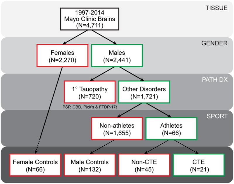
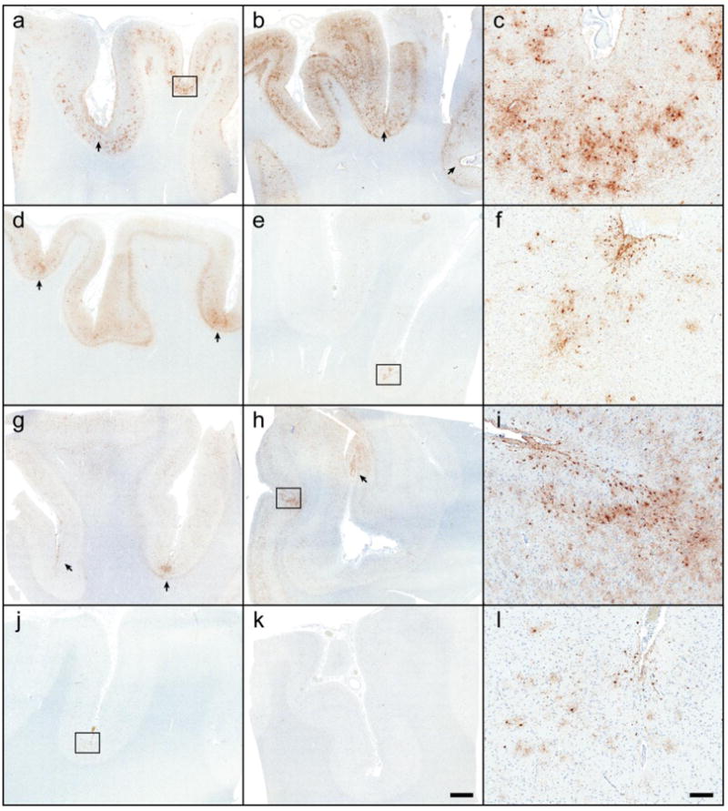
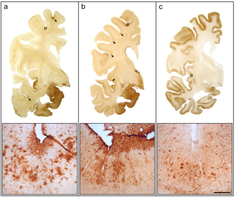
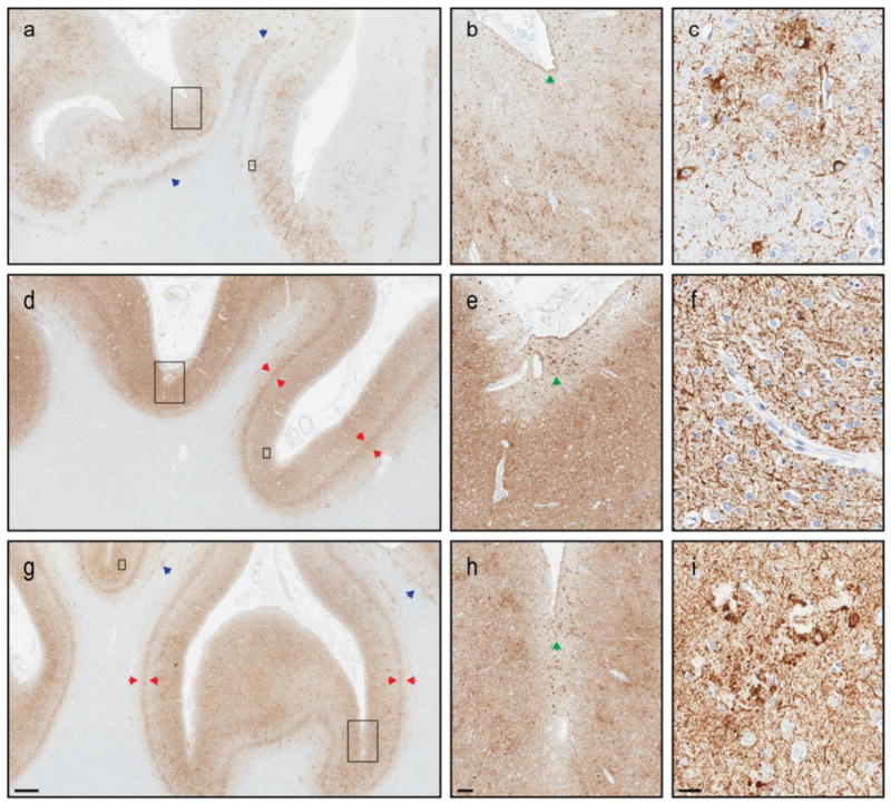
Chronic traumatic encephalopathy (CTE) is a progressive neurodegenerative disorder linked to repetitive traumatic brain injury (TBI) and characterized by deposition of hyperphosphorylated tau at the depths of sulci. We sought to determine the presence of CTE pathology in a brain bank for neurodegenerative disorders for individuals with and without a history of contact sports participation. Available medical records of 1721 men were reviewed for evidence of past history of injury or participation in contact sports. Subsequently, cerebral cortical samples were processed for tau immunohistochemistry in cases with a documented history of sports exposure as well as age- and disease-matched men and women without such exposure. For cases with available frozen tissue, genetic analysis was performed for variants in APOE, MAPT, and TMEM106B. Immunohistochemistry revealed 21 of 66 former athletes had cortical tau pathology consistent with CTE. CTE pathology was not detected in 198 individuals without exposure to contact sports, including 33 individuals with documented single-incident TBI sustained from falls, motor vehicle accidents, domestic violence, or assaults. Among those exposed to contact sports, those with CTE pathology did not differ from those without CTE pathology with respect to noted clinicopathologic features. There were no significant differences in genetic variants for those with CTE pathology, but we observed a slight increase in MAPT H1 haplotype, and there tended to be fewer homozygous carriers of the protective TMEM106B rs3173615 minor allele in those with sports exposure and CTE pathology compared to those without CTE pathology. In conclusion, this study has identified a small, yet significant, subset of individuals with neurodegenerative disorders and concomitant CTE pathology. CTE pathology was only detected in individuals with documented participation in contact sports. Exposure to contact sports was the greatest risk factor for CTE pathology. Future studies addressing clinical correlates of CTE pathology are needed.

Keywords: Brain bank; Chronic traumatic encephalopathy; Microtubule-associated protein tau; Sports; Traumatic brain injury.


Figures

Figure 1
Figure 1
Selection criteria for Mayo Clinic Florida cohort. There are 4,711 brains with available formalin-fixed tissue recruited between the years 1997 and 2014. Of these 4,711 brains, 2,441 were male and 1,721 males did not have a prior neuropathologic diagnosis (Path Dx) of a primary tauopathy, namely progressive supranuclear palsy (PSP), corticobasal degeneration (CBD), Pick's disease (Pick's), or frontotemporal lobar degeneration with parkinsonism with a known MAPT mutation (FTDP-17t). Available clinical records of these 1,721 males were queried for any description of involvement in a contact sport, and 66 individuals did have notation regarding former athletics participation. Screening cortical tissue via tau immunohistochemistry (dashed lines) identified 21 cases with CTE pathology and 45 cases that failed to meet study standards of CTE pathology. In addition, tau immunohistochemistry was also performed on 66 female and 132 male age- and disease-matched (Path Dx) controls without documentation of sports involvement.
Figure 2
Figure 2
Chronic traumatic encephalopathy in the Mayo Clinic Florida cohort. Tau immunohistochemistry on tissue from the frontal cortex (a, d, g, j) and parietal cortex (b, e, h, k) in 66 athletes and 198 controls revealed CTE pathology in 21 cases, including a former boxer (a-c: Case #20), a former high school and collegiate American football player (d-f: Case #12), and a multi-sport athlete (football, basketball and baseball)(g-i: Case #21). One case initially without clinical documentation of contact sports and CTE pathology was subsequently discovered to be a former high school football player (j-l: Case #4). Microscopically, CTE foci (arrows and boxes; c, f, i, l) were characterized by clustered glial and neuronal lesions primarily at the depths of sulci and surrounding vessels. Bar: 2 mm on large sections, 20 μm on insets.
Figure 3
Figure 3
Distribution of chronic traumatic encephalopathy foci in mixed neurodegenerative cases. In 3 former football players (ages 78, 79 and 81), cortical foci of CTE pathology are observed in the frontal and temporal cortices of two individuals, one with pathologic Alzheimer's disease and Lewy body disease (a: Case #16) and one with pathologic Lewy body disease (b: Case #14), but not in a third individual with pathologic Alzheimer's disease (c: Case #46). Distributed prominently at the depths of sulci, some foci are large and span the entirety of the cortical gray matter (a) while others lesions are discrete, concentrated at the subpial surface (b). Bar: 15 mm coronal images, 200 μm inset.
Figure 4
Figure 4
Neuropathologic differentiation of chronic traumatic encephalopathy and Alzheimer's disease. Tau pathology in the frontal cortex of 3 male amateur boxers, 1 with CTE (Case #11: a-c) and 2 without CTE (Case #29: d-f; Case #32: g-i). All 3 cases have concomitant Alzheimer's disease (Braak Stage 6, Thal Phase 5). Co-morbid CTE pathology is patchy (a) with marked prominence at the depth of cerebral sulci (b). CTE pathognomonic lesions include focal neurofibrillary tangles and astrocytic tau pathology adjacent to penetrating blood vessels (c). This contrasts the more uniform laminar distribution of Alzheimer tau pathology (d, g), particularly in layer 5 (red arrows). Similar to Case #11, Case #29 and #32 both show subpial thorn-shaped astrocytes (b, e, h; green arrows) at the depths of sulci. However, this often associated CTE feature is non-specific without additional perivascular pathology (f) or neuritic tau pathology associated with cerebral amyloid angiopathy (i). Tau-immunoreactive white matter astrocytes (blue arrows) are another pathologic feature commonly observed in CTE (a) which lack disease specificity (g). Ultimately, confluent tau pathology in cases with a high burden of Alzheimer pathology may prohibit a definitive CTE diagnosis. Large inset: b, e, h; small inset: c, f, i. Measure bar: 1mm a, d, g; 100 μm b, e, h; 25 μm c, f, i.

References

    1. Aoki N, Murray ME, Ogaki K, et al. Hippocampal sclerosis in Lewy body disease is a TDP-43 proteinopathy similar to FTLD-TDP Type A. Acta Neuropathol. 2015;129:53–64. - PMC - PubMed
    1. Braak H, Braak E. Neuropathological stageing of Alzheimer-related changes. Acta Neuropathologica. 1991;82:239–259. - PubMed
    1. Cantu RC, Mueller FO. The prevention of catastrophic head and spine injuries in high school and college sports. Br J Sports Med. 2009;43:981–986. - PubMed
    1. Corsellis JA, Bruton CJ, Freeman-Browne D. The aftermath of boxing. Psychological Medicine. 1973;3:270–303. - PubMed
    1. DeKosky ST, Blennow K, Ikonomovic MD, Gandy S. Acute and chronic traumatic encephalopathies: pathogenesis and biomarkers. Nature Reviews Neurology. 2013;9:192–200. - PMC - PubMed

Publication types

MeSH terms