. 2016 Feb;149(2):277-88.
doi: 10.1093/toxsci/kfv234. Epub 2015 Oct 30.

Elevated Lifetime Lead Exposure Impedes Osteoclast Activity and Produces an Increase in Bone Mass in Adolescent Mice

Affiliations

Elevated Lifetime Lead Exposure Impedes Osteoclast Activity and Produces an Increase in Bone Mass in Adolescent Mice

Eric E Beier et al. Toxicol Sci. 2016 Feb.

Abstract

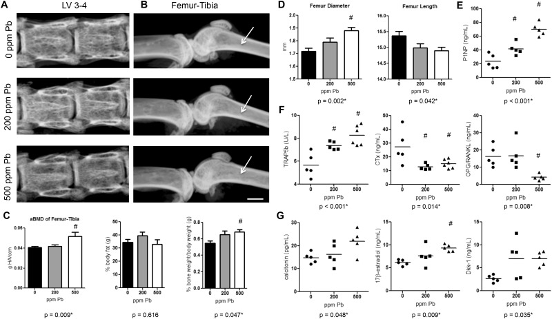
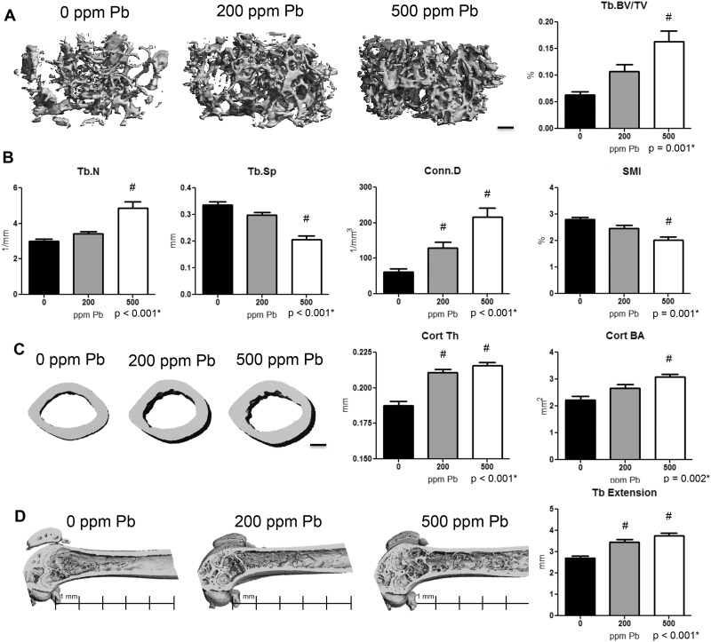
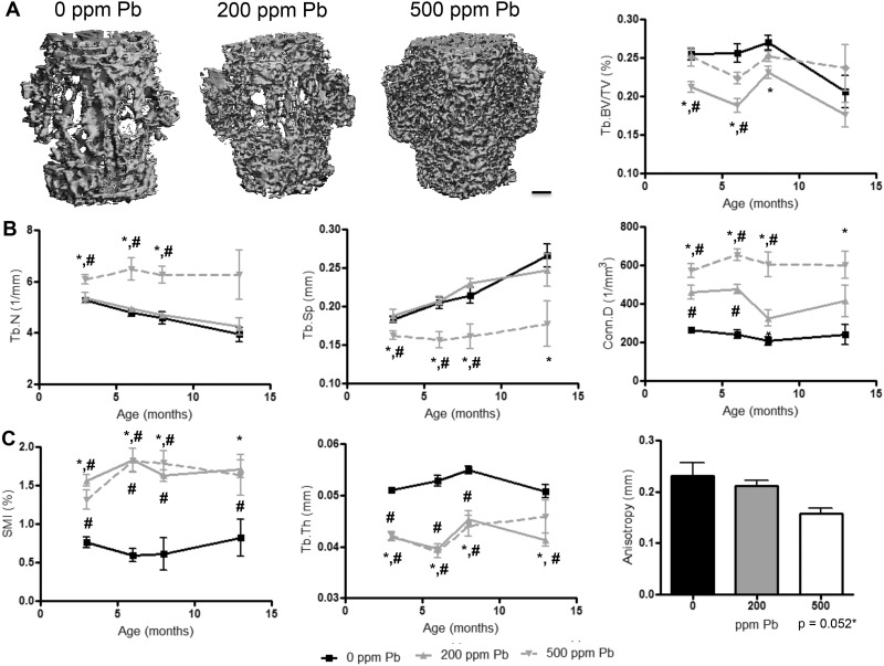
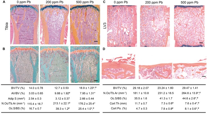
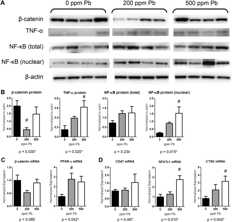
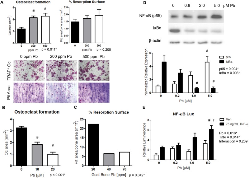
The heavy metal lead (Pb) has a deleterious effect on skeletal health. Because bone mass is maintained through a balance of bone formation and resorption, it is important to understand the effect of Pb levels on osteoblastic and osteoclastic activity. Pb exposure is associated with low bone mass in animal models and human populations; however, the correlation between Pb dosing and corresponding bone mass has been poorly explored. Thus, mice were exposed to increasing Pb and at higher levels (500 ppm), there was unexpectedly an increase in femur-tibial bone mass by 3 months of age. This is contrary to several studies alluded to earlier. Increased bone volume (BV) was accompanied by a significant increase in cortical thickness of the femur and trabecular bone that extended beyond the epiphyseal area into the marrow cavity. Subsequent evaluations revealed an increase in osteoclast numbers with high Pb exposure, but a deficiency in osteoclastic activity. These findings were substantiated by observed increases in levels of the resorption-altering hormones calcitonin and estrogen. In addition we found that pro-osteoclastic nuclear factor-kappa beta (NF-κB) pathway activity was dose dependently elevated with Pb, both in vivo and in vitro. However, the ability of osteoclasts to resorb bone was depressed in the presence of Pb in media and within test bone wafers. These findings indicate that exposure to high Pb levels disrupts early life bone accrual that may involve a disruption of osteoclast activity. This study accentuates the dose dependent variation in Pb exposure and consequent effects on skeletal health.

Keywords: NF-kappa B; Pb; bone density; bone turnover; osteoclast.


Figures

FIG. 1.
FIG. 1.
Elevated Pb exposure increases BMD, distorts bone growth, and alters serum bone biomarkers in female mice. Female C57Bl/6 mice were continuously exposed to 0, 200, or 500 ppm Pb through their drinking water. Radiographic images of vertebrae (A) and femur-tibia (B) were taken ex vivo of each treatment group at 3 months of age. Radiolucency in the trabecular region (arrows-B) highlight changes in trabecular architecture. Bar: 750 µm. C, DXA quantification of bone mass, bone weight, and body fat is shown. D, Femur diameter and length were measured to determine mouse stature at 6 months. Bone formation biomarkers (E), bone resorption indicators (F), and hormones and signaling molecules (G) were measured using standard ELISA methods at 3 months. Data represent mean ± SEM, n = 6 mice/group. Scatter plots are expressed as a population spread with an average bar (n = 5–6/group). *P < .05 for effect of Pb, #P < .05 for multiple comparisons versus 0 Pb.
FIG. 2.
FIG. 2.
Increased bone mass and cortical properties in female mice with elevated Pb exposure. A, Representative images (left) of transverse sections of the distal femur selected based on the median BV/TV (right) are presented at 6 months of age. B, Quantitative microCT determination of trabecular bone parameters and structure. C, Representative transverse sections of femur midshaft (left) and analysis of cortical parameters (right) including thickness and area are shown. D, Trabecular bone extension up the femoral midshaft was measured among groups, with a scale shown beneath representative images. Bars: 200 µm. Data represent mean ± SEM of 6 mice/group. *P < .05 for effect of Pb, #P < .05 for multiple comparisons versus 0 Pb.
FIG. 3.
FIG. 3.
Pb exposure impairs trabecular bone properties in the lumbar spine of female mice. A, Images are representative transverse sections of LV4 (left, bar: 200 µm) selected based on the median BV/TV (right) at 6 months. B, Additional microCT analyses of trabecular bone properties are provided of each group over time. C, Architectural measures of trabecular shape, thickness, and directionality are presented over time, and at 12 months for anisotropy. Data represent mean ± SEM of 5 mice/group. *P < .05 for effect of Pb, #P < .05 for multiple comparisons versus 0 Pb.
FIG. 4.
FIG. 4.
Pb exposure increases adipocyte formation and osteoclast formation, and vitiates cortical bone quality in female mice. At 6 months of age, bone specimens were stained with ABH (A, C, D) or TRAP (B). Representative images of trabecular (A, B, C) and cortical (D) bone of proximal tibias and lumbar vertebrae were selected based on the median BV/TV of each group (top). The effect of Pb on histologic parameters were calculated and displayed in the tables (bottom). Bar: (A–C) 100 µm, (D) 500 µm. Data represent mean ± SEM of 4 mice/group. *P < .05 for effect of Pb, #P < .05 for multiple comparisons versus 0 Pb.
FIG. 5.
FIG. 5.
Increased expression of osteogenic and adipogenic factors in bone from Pb-exposed female mice. At 6 months of age protein and RNA were isolated from tibial bone after aspiration of bone marrow cells. A, Western blots of β-catenin, TNF-α, NF-κB (total protein lysates and nuclear fraction), and β-actin are depicted for 4 samples from 0, 200, and 500 ppm Pb-treated mice and B, quantified using densitometry. C, β-catenin and PPAR-γ gene expression measured using RT-PCR. D, Osteoclastic genes CD47, NFATc1, and CTSK gene expression measured using RT-PCR. Data represent mean SEM (n = 4 mice/group). *P < .05 for effect of Pb, #P < .05 for multiple comparisons versus 0 Pb.
FIG. 6.
FIG. 6.
Pb promotes osteoclast formation but osteoclast activity is inhibited by elevated Pb exposure. Osteoclast formation and activity was assessed on bovine cortical bone wafers. A, Bone marrow cells isolated from Pb-exposed mice were evaluated for TRAP+ osteoclasts and pit resorption. B, Osteoclastic formation was evaluated from bone marrow cells treated with Pb though the media. C, Pb-intoxicated bone wafers were constructed from goats and were resistant to osteoclastic resorption. NF-κB signaling was determined by Western blot (D) and luciferase activity (E) in response to increasing Pb and recombinant TNF-α in RAW264.7 cells. Data represent mean ± SEM for 3 trials. *P < .05 for effect of variable, #P < .05 for multiple comparisons versus control, P < .05 for multiple comparisons versus 5 µM Pb.

References

    1. Ambrose T. M., Al-Lozi M., Scott M. G. (2000). Bone lead concentrations assessed by in vivo X-ray fluorescence. Clin. Chem. 46(8 Pt 1), 1171–1178. - PubMed
    1. Anderson J. M. (2000). Multinucleated giant cells. Curr. Opin. Hematol. 7, 40–47. - PubMed
    1. Arai F., Miyamoto T., Ohneda O., Inada T., Sudo T., Brasel K., Miyata T., Anderson D. M., Suda T. (1999). Commitment and differentiation of osteoclast precursor cells by the sequential expression of c-Fms and receptor activator of nuclear factor kappaB (RANK) receptors. J. Exp. Med. 190, 1741–1754. - PMC - PubMed
    1. Bagchi D., Preuss H. G. (2005). Effects of acute and chronic oval exposure of lead on blood pressure and bone mineral density in rats. J. Inorg. Biochem. 99, 1155–1164. - PubMed
    1. Ballew C., Khan L. K., Kaufmann R., Mokdad A., Miller D. T., Gunter E. W. (1999). Blood lead concentration and childrens anthropometric dimensions in the Third National Health and Nutrition Examination Survey (NHANES III), 1988-1994. J. Pediatrics 134, 623–630. - PubMed

Publication types