. 2016 Jan;170(1):263-72.
doi: 10.1104/pp.15.01529. Epub 2015 Oct 30.

In Vivo Quantification of Peroxisome Tethering to Chloroplasts in Tobacco Epidermal Cells Using Optical Tweezers

Affiliations

In Vivo Quantification of Peroxisome Tethering to Chloroplasts in Tobacco Epidermal Cells Using Optical Tweezers

Hongbo Gao et al. Plant Physiol. 2016 Jan.

Abstract

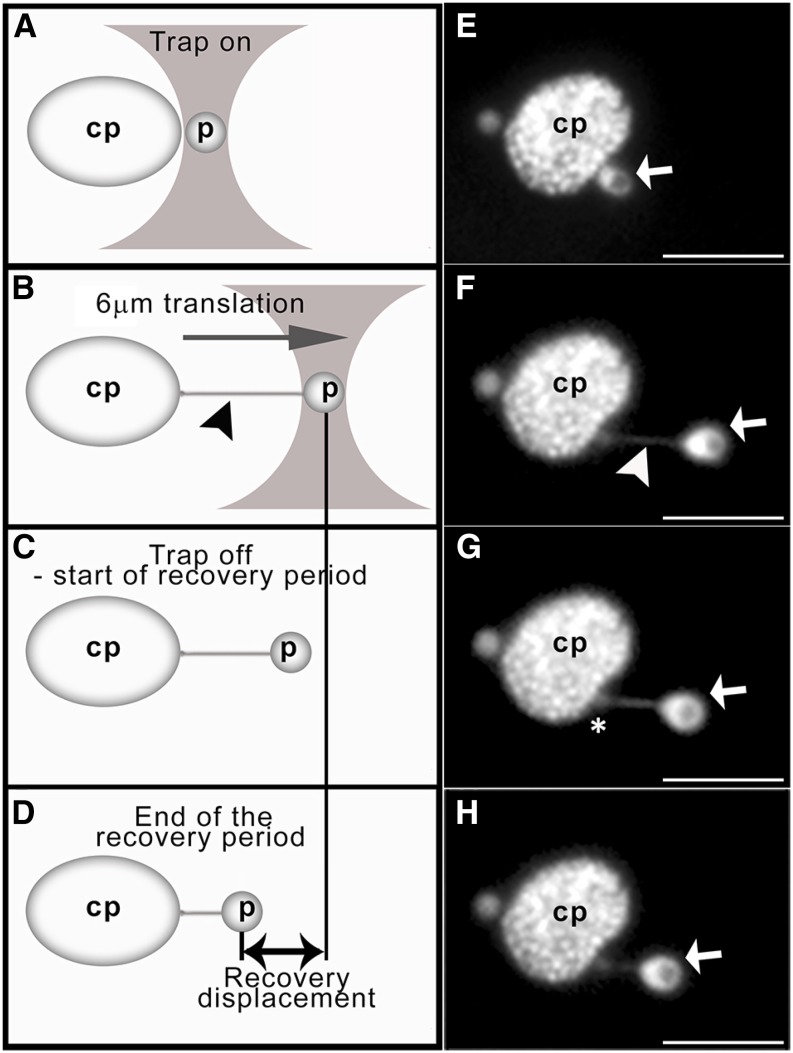
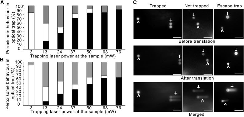
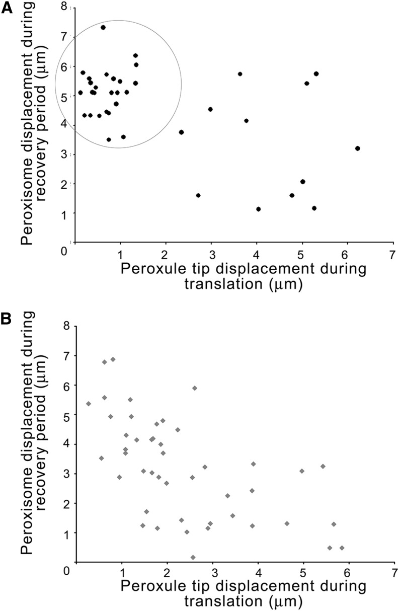
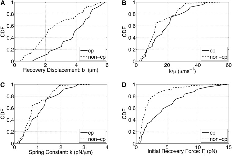
Peroxisomes are highly motile organelles that display a range of motions within a short time frame. In static snapshots, they can be juxtaposed to chloroplasts, which has led to the hypothesis that they are physically interacting. Here, using optical tweezers, we tested the dynamic physical interaction in vivo. Using near-infrared optical tweezers combined with TIRF microscopy, we were able to trap peroxisomes and approximate the forces involved in chloroplast association in vivo in tobacco (Nicotiana tabacum) and observed weaker tethering to additional unknown structures within the cell. We show that chloroplasts and peroxisomes are physically tethered through peroxules, a poorly described structure in plant cells. We suggest that peroxules have a novel role in maintaining peroxisome-organelle interactions in the dynamic environment. This could be important for fatty acid mobilization and photorespiration through the interaction with oil bodies and chloroplasts, highlighting a fundamentally important role for organelle interactions for essential biochemistry and physiological processes.


Figures

Figure 1.
Figure 1.
Optical trapping and movement of peroxisomes away from chloroplasts in tobacco leaf epidermal cells. Schematic representations of the trapping procedure (A–D) and corresponding micrographs (E–H) are shown. Upon turning the trap on (A and E) and moving the stage 6 μm at a set speed (B and F; referred to as the translation period), the trapped peroxisome (p; white arrows) is pulled away from the cp and a peroxule (arrowheads) is formed. Upon turning the trap off (C and G), the peroxisome recoils backs toward its original position next to the chloroplast (D and H). Peroxisome displacement during the recovery period (referred to as recovery displacement) is measured (double-headed arrow). The asterisk denotes the tip of the peroxule. Bars = 6 μm.
Figure 2.
Figure 2.
Higher optical trapping laser power is required to trap and move peroxisomes away from chloroplasts. The noncp (A) and cp (B) peroxisomes underwent the optical trapping protocol using various trapping laser powers, and their trapping characteristics were scored: peroxisomes that remained in the trap over the 6-μm translation (black bars), were unable to be trapped (white bars), or escaped the trap during the translation (gray bars). Percentages displayed are based on weighted means from a set of independent experiments. Supplemental Figure S1 compares cp with noncp peroxisomes for all three trapping categories and indicates significant differences between peroxisomes that are trapped or that escape from the trap for cp versus noncp samples. The relationship between optical laser trap power and peroxule formation is given in Supplemental Table S1. C shows still images from Supplemental Movie S3, C to E, representing before and after translation events for peroxisomes that are trapped, not trapped, or escape the trap during the translation event (arrowhead). Note that peroxisomes not subjected to trapping in the same cell are shown for comparison (arrows). The translation event is based on the movement of the stage and not the trap. Composite images of frames captured during the translation event show that the trapped peroxisome does not appear to move whereas organelles that escape the trap or are not trapped result in comet-like tails (merged). Bars = 6 μm.
Figure 3.
Figure 3.
Correlation between peroxisome displacement during the recovery period and peroxule tip displacement during translation indicates anchored tethering between chloroplasts and peroxisomes. Peroxule tip displacement during the translation period was plotted against the peroxisome displacement during the recovery period for cp (A; n = 37) and noncp (B; n = 46) peroxisomes. Peroxisomes were trapped with 37-mW optical trap laser power followed by a 21.5-s recovery period. The behavior of cp samples is indicative of anchored tethers, where the peroxule tip represents the base of the tether: small peroxule tip displacement combined with large peroxisome recovery displacement (circle). Note that the sample sizes are different from those in Supplemental Table S1, as displacement could only be measured if the peroxule was observable for the entire period.
Figure 4.
Figure 4.
Cumulative distribution functions (CDF) of model parameters for cp and noncp peroxisomes. Recovery displacement b is larger for cp than noncp peroxisomes (A), whereas k/μ values, indicative of the tether stiffness, are broadly similar (B). Also shown are the derived spring constants (C) and initial recovery forces (D) calculated assuming a viscosity of 0.06 Pa s for the cytoplasm. Values are derived using the spring model described in Supplemental Figures S2 to S4.

References

    1. Andersson MX, Goksör M, Sandelius AS (2007) Optical manipulation reveals strong attracting forces at membrane contact sites between endoplasmic reticulum and chloroplasts. J Biol Chem 282: 1170–1174 - PubMed
    1. Asada K. (2006) Production and scavenging of reactive oxygen species in chloroplasts and their functions. Plant Physiol 141: 391–396 - PMC - PubMed
    1. Avisar D, Prokhnevsky AI, Makarova KS, Koonin EV, Dolja VV (2008) Myosin XI-K is required for rapid trafficking of Golgi stacks, peroxisomes, and mitochondria in leaf cells of Nicotiana benthamiana. Plant Physiol 146: 1098–1108 - PMC - PubMed
    1. Barton K, Mathur N, Mathur J (2013) Simultaneous live-imaging of peroxisomes and the ER in plant cells suggests contiguity but no luminal continuity between the two organelles. Front Physiol 4: 196. - PMC - PubMed
    1. Brandizzi F, Snapp EL, Roberts AG, Lippincott-Schwartz J, Hawes C (2002) Membrane protein transport between the endoplasmic reticulum and the Golgi in tobacco leaves is energy dependent but cytoskeleton independent: evidence from selective photobleaching. Plant Cell 14: 1293–1309 - PMC - PubMed

Publication types

LinkOut - more resources