. 2016:1369:113-23.
doi: 10.1007/978-1-4939-3145-3_9.

Characterization of Septin Ultrastructure in Budding Yeast Using Electron Tomography

Affiliations

Characterization of Septin Ultrastructure in Budding Yeast Using Electron Tomography

Aurélie Bertin et al. Methods Mol Biol. 2016.

Abstract

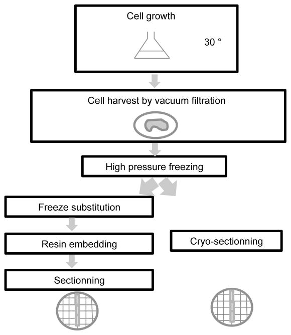
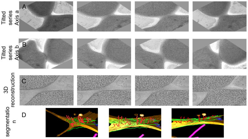
Septins are essential for the completion of cytokinesis. In budding yeast, Saccharomyces cerevisiae, septins are located at the bud neck during mitosis and are closely connected to the inner plasma membrane. In vitro, yeast septins have been shown to self-assemble into a variety of filamentous structures, including rods, paired filaments, bundles, and rings (Bertin et al. Proc Natl Acad Sci U S A, 105(24):8274-8279, 2008; Garcia et al. J Cell Biol, 195(6):993-1004, 2011; Bertin et al. J Mol Biol, 404(4):711-731, 2010). Using electron tomography of freeze-substituted sections and cryo-electron tomography of frozen sections, we determined the three-dimensional organization of the septin cytoskeleton in dividing budding yeast with molecular resolution (Bertin et al. Mol Biol Cell, 23(3):423-432, 2012; Bertin and Nogales. Commun Integr Biol 5(5):503-505, 2012). Here, we describe the detailed procedures used for our characterization of the septin cellular ultrastructure.

Keywords: Budding yeast; Cryo-sectio ning; Cryo-tomography; Cytokinesis; Image process ing; Septin.


Figures

Figure 1
Figure 1. Schematic of the procedure used for sample preparation for electron microscopy analysis
After cell growth, the cells are harvested by vacuum filtration and vitrified by high pressure freezing. Two strategies can then be employed. Either the vitrified cells are cryo-sectioned right before observation in the scope (or storage) or the cells are freeze substituted, embedded in resin at room temperature and sectioned.
Figure 2
Figure 2. Snapshots from a reconstructed tomogram and its corresponding model
Panel A. Images from a tilted series collected from a resin-embedded sample. Panel B. After the sample has been rotated by 90°, another tilted series was collected from which a few images are shown. Panel C. Images from different slices of the corresponding 3D reconstruction. Panel D. Model obtained after segmentation of the reconstructed tomogram.

References

    1. Bertin A, McMurray MA, Grob P, Park SS, Garcia G, 3rd, Patanwala I, Ng HL, Alber T, Thorner J, Nogales E. Saccharomyces cerevisiae septins: supramolecular organization of heterooligomers and the mechanism of filament assembly. Proc Natl Acad Sci U S A. 2008;105(24):8274–8279. - PMC - PubMed
    1. Garcia G, 3rd, Bertin A, Li Z, Song Y, McMurray MA, Thorner J, Nogales E. Subunit-dependent modulation of septin assembly: budding yeast septin Shs1 promotes ring and gauze formation. J Cell Biol. 2011;195(6):993–1004. doi: 10.1083/jcb.201107123. - DOI - PMC - PubMed
    1. Bertin A, McMurray MA, Thai L, Garcia G, 3rd, Votin V, Grob P, Allyn T, Thorner J, Nogales E. Phosphatidylinositol-4,5-bisphosphate promotes budding yeast septin filament assembly and organization. J Mol Biol. 2010;404(4):711–731. doi: 10.1016/j.jmb.2010.10.002. - DOI - PMC - PubMed
    1. Bertin A, McMurray MA, Pierson J, Thai L, McDonald KL, Zehr EA, Garcia G, 3rd, Peters P, Thorner J, Nogales E. Three-dimensional ultrastructure of the septin filament network in Saccharomyces cerevisiae. Mol Biol Cell. 2012;23(3):423–432. doi: 10.1091/mbc.E11-10-0850. - DOI - PMC - PubMed
    1. Bertin A, Nogales E. Septin filament organization in Saccharomyces cerevisiae. Commun Integr Biol. 2012;5(5):503–505. doi: 10.4161/cib.21125. - DOI - PMC - PubMed

Publication types

MeSH terms

LinkOut - more resources