Review
. 2015 Dec 2;43(21):10083-101.
doi: 10.1093/nar/gkv1136. Epub 2015 Oct 30.

The current state of eukaryotic DNA base damage and repair

Affiliations
Review

The current state of eukaryotic DNA base damage and repair

Nicholas C Bauer et al. Nucleic Acids Res. .

Abstract

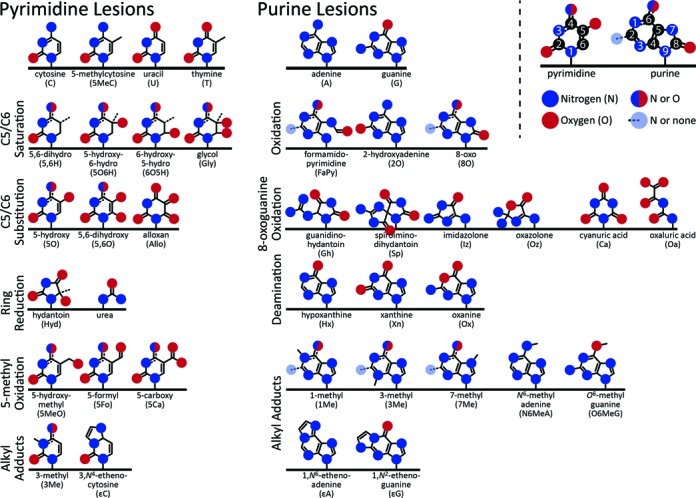
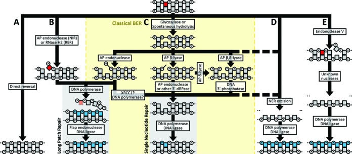
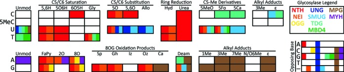
DNA damage is a natural hazard of life. The most common DNA lesions are base, sugar, and single-strand break damage resulting from oxidation, alkylation, deamination, and spontaneous hydrolysis. If left unrepaired, such lesions can become fixed in the genome as permanent mutations. Thus, evolution has led to the creation of several highly conserved, partially redundant pathways to repair or mitigate the effects of DNA base damage. The biochemical mechanisms of these pathways have been well characterized and the impact of this work was recently highlighted by the selection of Tomas Lindahl, Aziz Sancar and Paul Modrich as the recipients of the 2015 Nobel Prize in Chemistry for their seminal work in defining DNA repair pathways. However, how these repair pathways are regulated and interconnected is still being elucidated. This review focuses on the classical base excision repair and strand incision pathways in eukaryotes, considering both Saccharomyces cerevisiae and humans, and extends to some important questions and challenges facing the field of DNA base damage repair.


Figures

Figure 1.
Figure 1.
Common base lesions. The basic chemical structures of the common base lesions that occur in DNA grouped by type. The basic nucleotides are shown at the top, and lesions are displayed to indicate the same modification occurring to multiple bases. Hydrogens are omitted. IUPAC numbering for pyrimidines and purines is illustrated at the top right.
Figure 2.
Figure 2.
Base, sugar, and single-strand break lesion repair pathways. Base lesions can be processed by: (A) direct reversal; (B) nucleotide incision repair (NIR), ribonucleotide excision repair (RER); (C) classical base excision repair (BER); (D) nucleotide excision repair (NER) or (E) endonuclease Vmediated excision repair. Within the BER pathway (C), multiple semi-redundant pathways are available for processing abasic sites. Pathway choice depends on the lesion, and some lesions can be acted on by multiple pathways. Squares (nucleosides) and connected circles (phosphates) represent a section of a DNA molecule, with the base lesion indicated in red. De novo synthesized bases are shown in blue. The enzyme activity responsible for each step is noted. The dashed segments indicate a fallback pathway. AP = apurinic/apyrimidinic site; dRP = deoxyribose phosphate.
Figure 3.
Figure 3.
Substrate specificities of base excision repair glycosylases. Each square represents a DNA base lesion, with the row indicating the original base and the column indicating the particular lesion, grouped by shared features. ‘Unmod’ = unmodified. Refer to Figure 1 for lesion abbreviations. Coloration within each box indicates the glycosylase families that recognize the lesion and excise it from DNA, according to the legend at the top-right. The matrix at the bottom-right illustrates the specificity of the glycosylase families with respect to the base opposite from a lesion; — indicates single-stranded DNA. Note that this diagram does not account for differences in enzyme kinetics between or within families. Example: OGG (yellow) excises guanine-derived formamidopyrimidine, 8-oxoadenine, and 8-oxoguanine when these lesions are opposite to cytosine.

References

    1. Bjelland S., Seeberg E. Mutagenicity, toxicity and repair of DNA base damage induced by oxidation. Mutat. Res. 2003;531:37–80. - PubMed
    1. Morreall J.F., Petrova L., Doetsch P.W. Transcriptional mutagenesis and its potential roles in the etiology of cancer and bacterial antibiotic resistance. J. Cell. Physiol. 2013;228:2257–2261. - PMC - PubMed
    1. Lindahl T. Instability and decay of the primary structure of DNA. Nature. 1993;362:709–715. - PubMed
    1. Brown E.J., Baltimore D. ATR disruption leads to chromosomal fragmentation and early embryonic lethality. Genes Dev. 2000;14:397–402. - PMC - PubMed
    1. Vermeij W.P., Hoeijmakers J.H.J., Pothof J. Aging: not all DNA damage is equal. Curr. Opin. Genet. Dev. 2014;26:124–130. - PubMed

Publication types

Substances