. 2015 Dec 1;195(11):5318-26.
doi: 10.4049/jimmunol.1500559. Epub 2015 Oct 30.

β-Glucan-Activated Human B Lymphocytes Participate in Innate Immune Responses by Releasing Proinflammatory Cytokines and Stimulating Neutrophil Chemotaxis

Affiliations

β-Glucan-Activated Human B Lymphocytes Participate in Innate Immune Responses by Releasing Proinflammatory Cytokines and Stimulating Neutrophil Chemotaxis

Mohamed F Ali et al. J Immunol. .

Abstract

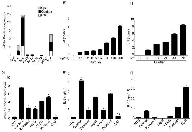
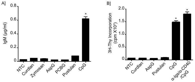
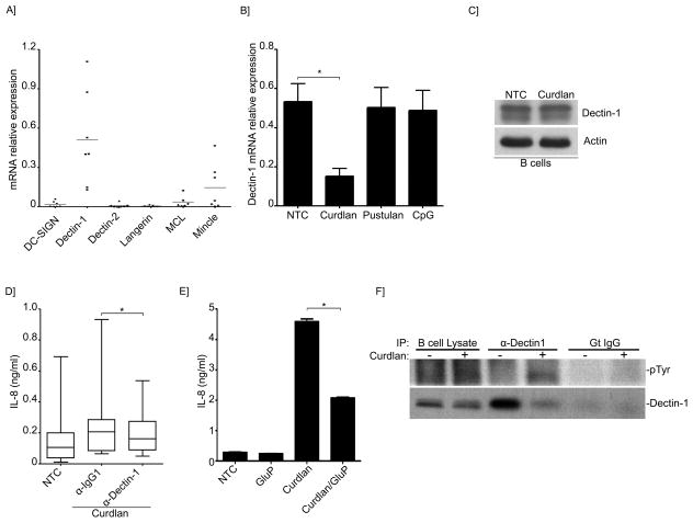
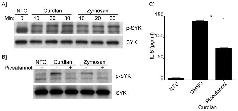
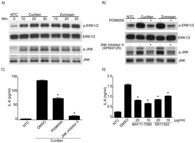
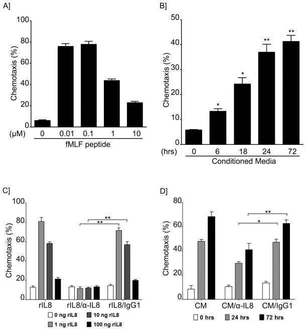
B lymphocytes play an essential regulatory role in the adaptive immune response through Ab production during infection. A less known function of B lymphocytes is their ability to respond directly to infectious Ags through stimulation of pattern recognition receptors expressed on their surfaces. β-Glucans are carbohydrates present in the cell wall of many pathogenic fungi that can be detected in the peripheral blood of patients during infection. They have been shown to participate in the innate inflammatory response, as they can directly activate peripheral macrophages and dendritic cells. However, their effect as direct stimulators of B lymphocytes has not been yet fully elucidated. The aim of this study was to examine the molecular mechanisms and cytokine profiles generated following β-glucan stimulation of B lymphocytes, compared with the well-established TLR-9 agonist CpG oligodeoxynucleotide (CpG), and study the participation of β-glucan-stimulated B cells in the innate immune response. In this article, we demonstrate that β-glucan-activated B lymphocytes upregulate proinflammatory cytokines (TNF-α, IL-6, and IL-8). Of interest, β-glucan, unlike CpG, had no effect on B lymphocyte proliferation or IgM production. When compared with CpG (TLR9 agonist), β-glucan-activated cells secreted significantly higher levels of IL-8. Furthermore, IL-8 secretion was partially mediated by Dectin-1 and required SYK, MAPKs, and the transcription factors NF-κB and AP-1. Moreover, we observed that conditioned media from β-glucan-stimulated B lymphocytes elicited neutrophil chemotaxis. These studies suggest that β-glucan-activated B lymphocytes have an important and novel role in fungal innate immune responses.


Figures

Figure 1
Figure 1. Cytokine profile from β-glucan stimulated human peripheral B-lymphocytes differs from CpG stimulated cells
(A) Quantitative real-time PCR for indicated mRNAs in non-treated cells (NTC) or B-cells stimulated with CpG or Curdlan. Stacked bars represent the relative expression of the cytokines tested after specific stimuli of B-lymphocytes as indicated. Segments of each bar are not additive. Expression was normalized to GAPDH. (B) IL-8 was measured by ELISA in the cell supernatant after Curdlan stimulation at the indicated concentrations or (C) after stimulation with Curdlan for the indicated period of time. (D) Quantitative real-time PCR for IL-8 mRNAs in NTC or B-cells stimulated with the different β-glucan preparations or CpG. Expression was normalized to GAPDH. (E) IL-8 and (F) IL-10 were measured by ELISA in the supernatants of NTC, after stimulation with different β-glucan preparations or CpG as indicated. Shown are data from a representative experimental run, typical of at least three separate experimental preparations, performed with different B-cell preparations. *p<0.0001 when compared with the NTC.
Figure 2
Figure 2. Lack of IgM production and B-cell proliferation after β-glucan stimulation
(A) IgM was measured by ELISA in the supernatants of B-lymphocytes from NTC, treated with different β-glucan preparations or CpG as indicated for a total of 5 days. (B) 3H-Thymidine incorporation was assessed in NTC, stimulated cells with different β-glucan preparations, CpG or Immunoglobulin cocktail control. Shown are data from a representative experimental run, typical of at least three separate experimental preparations, performed with different B-cell preparations. *p<0.0001 when compared with the NTC.
Figure 3
Figure 3. Dectin-1 participates in IL-8 secretion induced by β-glucans
(A) Quantitative real-time PCR for the indicated C-lectin receptors mRNAs in B-lymphocytes. Expression was normalized to GAPDH. Shown are the data from 7 different B-cell preparations. (B) Quantitative real-time PCR for Dectin-1 mRNAs in B-lymphocytes of NTC, or after stimulation with Curdlan, pustulan or CpG. Expression was normalized to GAPDH. Shown are the data from 3 different B-cell preparations. *p< 0.05 (C) Immunoblot analysis of Dectin-1 in unstimulated or Curdlan-stimulated B-lymphocytes. (D)IL-8 was measured by ELISA in the supernatant of NTC and after Curdlan stimulation. Cells were pretreated with Dectin-1 neutralizing antibodies or isotype control as indicated. Data were analyzed by the non-parametric Wilcoxon matched-pairs signed rank test to compare the medians of 5 different B-cell preparations. (E) Secretion of IL-8 into the cell supernatant was measured by ELISA of NTC or Curdlan-stimulated cells. Cells were pretreated with glucan phosphate as indicated. Shown is a representative run of 3 independent experiments performed with 3 different B-cell preparations. *p< 0.01. (F) Endogenous Dectin-1 was immunoprecipitated from unstimulated and Curdlan-stimulated cells phospho-tyrosine antibody was used to detect Dectin-1 phosphorylation in the immunoprecipitates. Total Dectin-1 was used as a control.
Figure 4
Figure 4. IL-8 secretion does not require internalization of β-glucan particles in B-lymphocytes
Fluorescently labeled zymosan particles were incubated for the indicated periods of time with B-lymphocytes or RAW cells. Internalization was assessed by confocal fluorescence microscopy. Bar = 5 μm.
Figure 5
Figure 5. Syk activation and participation in IL-8 secretion induced by β-glucans
(A) Immunoblot analysis of phospho- and total-Syk in unstimulated and stimulated cells with Curdlan or Zymosan as indicated; (B) for some conditions cells were pretreated with piceatannol as indicated. (C) IL-8 was measured by ELISA in the cell supernatant of NTC and after Curdlan stimulation. Cells were pretreated with piceatannol or vehicle control as indicated. *p<0.0001. Shown are data from a representative experimental run, typical of at least three separate experimental preparations, performed with different B-cell preparations.
Figure 6
Figure 6. MAPKs (ERK and JNK) and the transcription factors AP-1 and NF-κB participate in IL-8 secretion
(A) Immunoblot analysis of ERK and JNK (phosphorylated and total) in NTC and stimulated cells with Curdlan or Zymosan as indicated; (B) for some conditions cells were pretreated with PD98059 (ERK inhibitor) or JNK inhibitor II as indicated. (C) IL-8 concentration was measured by ELISA in the cell supernatant of NTC and after Curdlan stimulation. Cells were pretreated with PD98059 and JNK inhibitor II or (D) BAY11 (NF-κB inhibitor), SR11 (AP-1 inhibitor) as indicated. DMSO was used as the vehicle control. Shown are data from a representative experimental run, typical of at least three separate experimental preparations, performed with different B-cell preparations. *p<0.0001 when compared with DMSO control.
Figure 7
Figure 7. Conditioned media from β-Glucan-activated B-lymphocytes induced neutrophil chemotaxis and is partially mediated by IL-8
Transwell chemotaxis assay of peripheral neutrophils towards different concentrations of (A) fMLF peptide (positive control) or (B) towards conditioned media form β-glucan treated B-lymphocytes for the indicated periods of time. Shown are data from a representative experimental run, typical of at least three separate experimental preparations, performed with different B-cell preparations. *p<0.05 and **p<0.01 when compared with NTC. Transwell chemotaxis assay of peripheral neutrophils towards different concentrations of recombinant IL-8 (C) or conditioned media (D) from β-glucan treated B cells in the presence or absence of anti-IL-8 or isotype control. *p<0.001 and **p<0.0001. Shown are data from a representative experimental run, typical of at least three separate experiments performed with different B cell preparations.

References

    1. Gerondakis S, Grumont RJ, Banerjee A. Regulating B-cell activation and survival in response to TLR signals. Immunol Cell Biol. 2007;85:471–475. - PubMed
    1. Meyer-Bahlburg A, Rawlings DJ. Differential impact of Toll-like receptor signaling on distinct B cell subpopulations. Front Biosci (Landmark Ed) 2012;17:1499–1516. - PMC - PubMed
    1. Fillatreau S. Cytokine-producing B cells as regulators of pathogenic and protective immune responses. Ann Rheum Dis. 2013;72:ii80–84. - PubMed
    1. Yehudai D, Snir A, Peri R, Halasz K, Haj T, Odeh M, Kessel A. B cell-activating factor enhances interleukin-6 and interleukin-10 production by ODN-activated human B cells. Scand J Immunol. 2012;76:371–377. - PubMed
    1. Klinman DM, Yi AK, Beaucage SL, Conover J, Krieg AM. CpG motifs present in bacteria DNA rapidly induce lymphocytes to secrete interleukin 6, interleukin 12, and interferon gamma. Proc Natl Acad Sci U S A. 1996;93:2879–2883. - PMC - PubMed

Publication types

MeSH terms