Review
. 2015 Dec;25(12):780-792.
doi: 10.1016/j.tcb.2015.09.005. Epub 2015 Oct 28.

When Physics Takes Over: BAR Proteins and Membrane Curvature

Affiliations
Review

When Physics Takes Over: BAR Proteins and Membrane Curvature

Mijo Simunovic et al. Trends Cell Biol. 2015 Dec.

Abstract

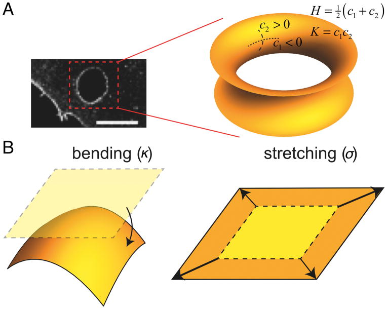
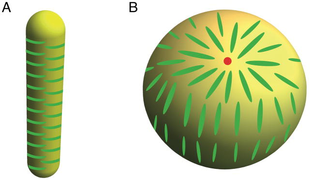
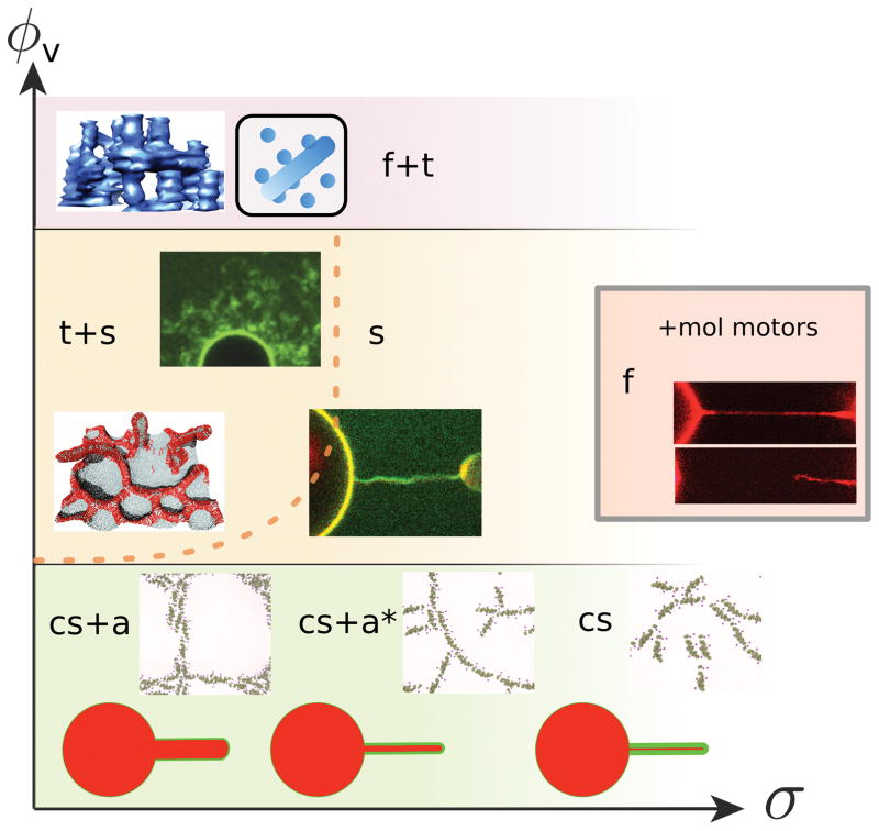
Cell membranes become highly curved during membrane trafficking, cytokinesis, infection, immune response, or cell motion. Bin/amphiphysin/Rvs (BAR) domain proteins with their intrinsically curved and anisotropic shape are involved in many of these processes, but with a large spectrum of modes of action. In vitro experiments and multiscale computer simulations have contributed in identifying a minimal set of physical parameters, namely protein density on the membrane, membrane tension, and membrane shape, that control how bound BAR domain proteins behave on the membrane. In this review, we summarize the multifaceted coupling of BAR proteins to membrane mechanics and propose a simple phase diagram that recapitulates the effects of these parameters.

Keywords: BAR proteins; curvature sensing; membrane curvature; membrane scission; membrane shaping; protein scaffold.


Figures

Figure i
Figure i
Elasticity and curvature of the membrane. (A) An example of a highly curved membrane at the trans-cellular tunnel (scale bar: 5 μm, adapted from [43]), displaying a negative Gaussian or saddle-like curvature. (B) Basic elastic membrane deformations: bending (characterized by bending stiffness, κ) and stretching (characterized by membrane tension, σ)
Figure ii
Figure ii
Common methods to study curvature-coupled proteins. (A) Micropipette aspiration of giant vesicles and extrusion of membrane tubules by optical tweezers. (B) Tethered small vesicles. (C) Coarse-grained model of a lipid vesicle decorated with N-BAR proteins (magnified in inset).
Figure 1
Figure 1. The structure and phenotype of BAR proteins
(A) Representative members of the BAR protein family, each subunit differently colored. Shown are (top) endophilin A2 (red-colored N-terminal amphipathic helices were added by hand), (center) FCHo2, (bottom) IRSp53. Dashed lines are guides for the eyes showing the intrinsic curvature of the proteins. (B) Binding of BAR proteins induces membrane tubulation. (Left) overexpression of β2 centaurin (BAR), scale bar: 20 μm, adapted from [4], reprinted with permission from AAAS; (top right) incubation of giant unilamellar vesicles (composition DOPC:DOPS:DOPE = 1:1:1, molar ratio) with fluorescently labeled (green) 1 μM amphiphysin 1 (N-BAR), scale bar: 5 μm, adapted from [20]; (bottom right) recruitment of amphiphysin 1 (N-BAR) to the plasma membrane by a chemical trigger (initiated at time zero), scale bar: 2 μm, adapted by permission from Macmillan Publishers Ltd: Scientific Reports [19], copyright (2014).
Figure 2
Figure 2. The biological role of BAR proteins
Shown is a cartoon of a cell (not to scale) with a non-comprehensive list of BAR proteins found in various curvature-related phenomena. Counter-clockwise from top left: IRSp53 and other I-BAR proteins colocalize with filopodia. Fluorescence image shows an enrichment of fluorescently labeled (green) C. elegans I-BAR domain on filopodia, scale bar: 5 μm, adapted from [24], with permission from Elsevier, copyright (2009). MIM (I-BAR) is found enriched on the edges of trans-cellular tunnels formed by bacterial toxins. Image shows a tunnel with fluorescently labeled MIM, scale bar: 5 μm, adapted from [43], with permission from Elsevier, copyright (2011). Amphiphysin 2 (N-BAR) is crucial for the formation of T-tubules (tubular invaginations in the membrane of skeletal and cardiac muscles). Image shows the localization of fluorescently labeled endogenous amphiphysin 2 on differentiated myotubes, scale bar: 10 μm. Adapted from [11], reprinted with permission from AAAS. Endophilin B1 (N-BAR) is key for the formation of reticular membrane morphology of the mitochondrion. Shown is a mitochondrial network stained with anti-endophilin B1 antibody. Adapted from [53], copyright (2004) by The Journal of Cell Biology. A variety of BAR proteins colocalize with endocytosis, e.g. FCHo2, Syp1, Bzz1 (F-BARs) are found at early stages of endocytosis, syndapin (F-BAR), various amphiphysins, endophilins (N-BARs), and sorting nexin 9 (N-BAR-like protein) were found at later stages of endocytosis. Electron microscopy image shows an ultrastructure of a membrane invagination in the course of clathrin-mediated endocytosis in yeast. Scale bar: 100 nm. Adapted from [56], with permission from Elsevier, copyright (2012). Many sorting nexins (N-BARs) are found on endosomes. Shown are structures of sorting nexins 1 (top) and 9 (bottom). Image shows a membrane tubule budding from an endosome coated by fluorescently labeled sorting nexin 1. Scale bar: 10 μm. Republished with permission of the Company of Biologists Ltd., from [58], permission conveyed through Copyright Clearance Center, Inc.
Figure 3
Figure 3. Influence of membrane geometry on BAR domain protein packing
(A) Anisotropic, rod-shape proteins pack efficiently on a cylindrical membrane. (B) On a spherical vesicle, there are necessarily at least two aster-like packing defects (indicated by a red dot).
Figure 4
Figure 4. Phase diagram illustrating the coupling between BAR protein density and membrane mechanics
Qualitatively different behaviors are found depending on position of the phase space spanned by the protein density, ϕv, on the membrane reservoir and the membrane tension, σ. The phase boundaries are schematic and their position will change depending on the intrinsic curvature of the protein, p. The different phases correspond to different behaviors protein-membrane system: cs = curvature sorting, a = linear assembly and netting, a* = disrupted linear assembly, t = tubulation, s = scaffolding, f = fission. The dashed line approximates the tension-inhibited onset of tubulation observed in [50]. The grey-framed inset represents high-density behavior of BAR proteins with added molecular-motor activity [9]. Image in f+t phase reprinted from [51], with permission from Elsevier, top image in t+s phase adapted from [20], bottom image in t+s kindly provided by Hiroshi Noguchi, image in s phase generated in our group, showing the stabilization of a membrane tubule by a BAR protein, as in [9, 20], method described in Box 2, image in f phase adapted from [9], images in the lowest-density part of the phase diagram, showing N-BAR protein assembly on a flat membrane, adapted from [26].

References

    1. McMahon HT, Gallop JL. Membrane curvature and mechanisms of dynamic cell membrane remodelling. Nature. 2005;438:590–596. - PubMed
    1. Atkins P, de Paula J. Atkins’ Physical Chemistry. OUP; Oxford: 2010.
    1. Goetz R, et al. Mobility and elasticity of self-assembled membranes. Phys Rev Lett. 1999;82:221–224.
    1. Peter BJ, et al. BAR domains as sensors of membrane curvature: the amphiphysin BAR structure. Science. 2004;303:495–499. - PubMed
    1. Saheki Y, De Camilli P. Synaptic vesicle endocytosis. Cold Spring Harb Perspect Biol. 2012;4:a005645. - PMC - PubMed

Publication types

LinkOut - more resources