Review
. 2015 Nov;100(11):1388-95.
doi: 10.3324/haematol.2015.128538.

Complement inhibitors to treat IgM-mediated autoimmune hemolysis

Affiliations
Review

Complement inhibitors to treat IgM-mediated autoimmune hemolysis

Diana Wouters et al. Haematologica. 2015 Nov.

Abstract

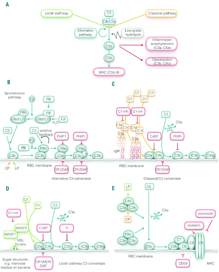
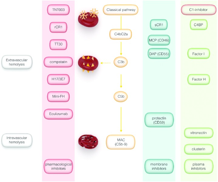
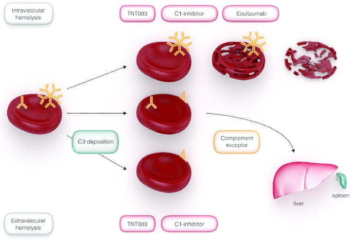
Complement activation in autoimmune hemolytic anemia may exacerbate extravascular hemolysis and may occasionally result in intravascular hemolysis. IgM autoantibodies as characteristically found in cold autoantibody autoimmune hemolytic anemia, in cold agglutinin disease but also in a considerable percentage of patients with warm autoantibodies are very likely to activate complement in vivo. Therapy of IgM-mediated autoimmune hemolytic anemia mainly aims to decrease autoantibody production. However, most of these treatments require time to become effective and will not stop immediate ongoing complement-mediated hemolysis nor prevent hemolysis of transfused red blood cells. Therefore pharmacological inhibition of the complement system might be a suitable approach to halt or at least attenuate ongoing hemolysis and improve the recovery of red blood cell transfusion in autoimmune hemolytic anemia. In recent years, several complement inhibitors have become available in the clinic, some of them with proven efficacy in autoimmune hemolytic anemia. In the present review, we give a short introduction on the pathogenesis of autoimmune hemolytic anemia, followed by an overview on the complement system with a special focus on its regulation. Finally, we will discuss complement inhibitors with regard to their potential efficacy to halt or attenuate hemolysis in complement-mediated autoimmune hemolytic anemia.


Figures

Figure 1.
Figure 1.
Overview of the complement system. (A) Overview of the complement system including the main activation pathways. (B) The alternative pathway is initiated by spontaneous low-grade conversion of C3 into active C3 (C3b), which together with activated factor B (Bb) forms the alternative C3 convertase which can induce additional C3 cleavage in a positive feedback loop. (C) The classical pathway is activated by antibodies [one IgM molecule, multiple (preferably 6) IgG molecules] leading to the formation of the classical C3 convertase (C2aC4b) by the activation C2 and C4 by C1s/C1r. (D) The lectin pathway is initiated by binding of MBL (or ficolins) to sugar structures followed by activation of C2 and C4 by MASP1/MASP2, leading to the formation of lectin C3 convertase (C2aC4b). (E) C3-activation by the classical, lectin or alternative C3 convertase results in the formation of the C5 convertase. C5 convertase subsequently activates C5 resulting in the formation of the membrane attack complex (MAC). C: complement factor; MAC: membrane attack complex; MBL: mannan binding lectin; MASP: MBL–associated serine protease; P: properdin; C1-inh: C1-inhibitor; FI: factor I; CR1: complement receptor 1; MCP: membrane co-factor protein; DAF: decay accelerating factor; C4BP: C4-binding protein; FH: factor H.
Figure 2.
Figure 2.
Regulation of the complement system by endogenous and pharmacological inhibitors. Plasmatic (green), membrane-bound (purple) and pharmaceutical (red) complement inhibitors. IgM is shown as pentamers on the red blood cells. C3b deposition is depicted as triangles on the red blood cell. Hemolysis by the MAC is depicted as red pores on the red blood cell membrane and a disintegrated membrane (so called red blood cell “ghost”). C4BP: C4b binding protein; MCP: membrane co-factor protein; DAF: decay accelerating factor; (s)CR1: soluble complement receptor 1; for other details see text.
Figure 3.
Figure 3.
Inhibition of intra- and extravascular hemolysis in AIHA by complement inhibitors. Complement-mediated removal of RBCs can occur intravascular or extravascular by complement-receptor mediated phagocytosis, mainly in the liver and to some extent also in the spleen. C3b deposition is depicted as triangles on the red blood cell. C5b deposition is depicted as circles on the red blood cell. Hemolysis by the MAC is depicted as a disintegrated membrane. Eculizumab inhibits intravascular hemolysis, whereas TNT003 and C1-inhibitor are able to inhibit both intravascular- and extravascular hemolysis (adapted from Zeerleder 3).

References

    1. Packman CH. Hemolytic anemia due to warm autoantibodies. Blood Rev 2008;22 (1):17–31. - PubMed
    1. Petz LD. Cold antibody autoimmune hemolytic anemias. Blood Rev 2008; 22(1): 1–15. - PubMed
    1. Zeerleder S. Autoimmune haemolytic anaemia - a practical guide to cope with a diagnostic and therapeutic challenge. Neth J Med 2011;69(4):177–184. - PubMed
    1. Barcellini W, Fattizzo B, Zaninoni A, et al. Clinical heterogeneity and predictors of outcome in primary autoimmune hemolytic anemia: a GIMEMA study of 308 patients. Blood 2014;124(19):2930–2936. - PubMed
    1. Lechner K, Jager U. How I treat autoimmune hemolytic anemias in adults. Blood 2010;116(11):1831–1838. - PubMed

MeSH terms

LinkOut - more resources